

Assessment of hemorrhage in pituitary macroadenoma by T2*-weighted gradient-echo MR imaging
- PMID: 17898201
- PMCID: PMC8134270
- DOI: 10.3174/ajnr.A0692
Assessment of hemorrhage in pituitary macroadenoma by T2*-weighted gradient-echo MR imaging
Abstract
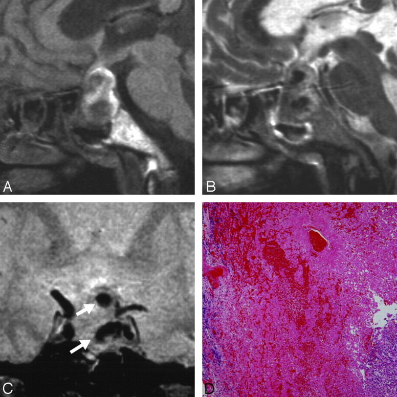
Background and purpose: Intratumoral hemorrhage occurs frequently in pituitary macroadenoma and manifests as pituitary apoplexy and recent or old silent hemorrhage. T2*-weighted gradient-echo (GE) MR imaging is the most sensitive sequence for the detection of acute and old intracranial hemorrhage. T2*-weighted GE MR imaging was used to investigate intratumoral hemorrhage in pituitary macroadenomas.
Materials and methods: Twenty-five consecutive patients who underwent total or subtotal resection of pituitary macroadenoma with heights from 17 to 53 mm, including 1 patient with classic pituitary apoplexy, underwent MR imaging before surgery, including T2*-weighted GE MR imaging. For histologic assessment of the hemorrhage in whole surgical specimens, we used hematoxylin-eosin staining.
Results: T2*-weighted GE MR imaging detected various types of dark lesions, such as "rim," "mass," "spot," and "diffuse" and combinations, indicating clinical and subclinical intratumoral hemorrhage in 12 of the 25 patients. The presence of intratumoral dark lesions on T2*-weighted GE MR imaging correlated significantly with the hemorrhagic findings on T1- and T2-weighted MR imaging (P < .02 and <.01, respectively), and the surgical and histologic hemorrhagic findings (P < .001 and <.001, respectively).
Conclusion: T2*-weighted GE MR imaging could detect intratumoral hemorrhage in pituitary adenomas as various dark appearances. Therefore, this technique might be useful for the assessment of recent and old intratumoral hemorrhagic events in patients with pituitary macroadenomas.
Figures


References
-
- Wakai S, Yamakawa K, Manaka S, et al. Spontaneous intracranial hemorrhage caused by brain tumor: its incidence and clinical significance. Neurosurgery 1982;10:437–44 - PubMed
-
- Mohr G, Hardy J. Hemorrhage, necrosis, and apoplexy in pituitary adenomas. Surg Neurol 1982;18:181–89 - PubMed
-
- Kyle CA, Laster RA, Burton EM, et al. Subacute pituitary apoplexy: MR and CT appearance. J Comput Assist Tomogr 1990;14:40–44 - PubMed
-
- Piotin M, Tampieri D, Rüfenacht DA, et al. The various MRI patterns of pituitary apoplexy. Eur Radiol 1999;9:918–23 - PubMed
-
- Lubina A, Olchovsky D, Berezin M, et al. Management of pituitary apoplexy: clinical experience with 40 patients. Acta Neurochir (Wien) 2005;147:151–57; discussion 157 - PubMed
Publication types
MeSH terms
LinkOut - more resources
Full Text Sources
Medical