. 2007 Sep 26;27(39):10365-71.
doi: 10.1523/JNEUROSCI.0630-07.2007.

In vitro characterization of Pittsburgh compound-B binding to Lewy bodies

Affiliations

In vitro characterization of Pittsburgh compound-B binding to Lewy bodies

Michelle T Fodero-Tavoletti et al. J Neurosci. .

Abstract

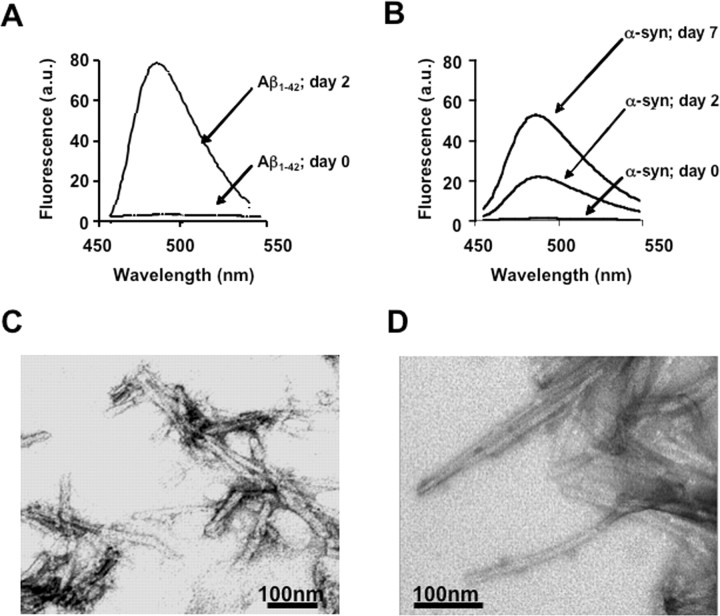
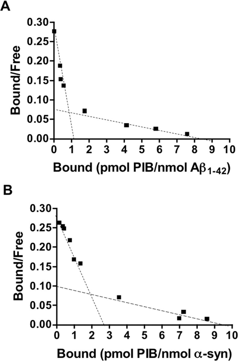
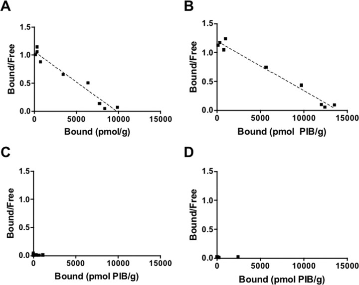
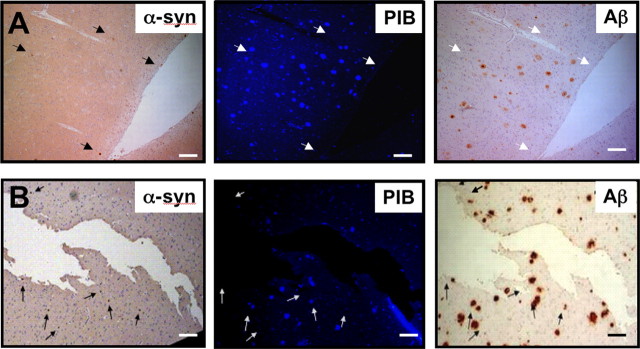
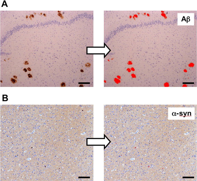
Dementia with Lewy bodies (DLB) is pathologically characterized by the presence of alpha-synuclein-containing Lewy bodies within the neocortical, limbic, and paralimbic regions. Like Alzheimer's disease (AD), Abeta plaques are also present in most DLB cases. The contribution of Abeta to the development of DLB is unclear. [11C]-Pittsburgh compound B ([11C]-PIB) is a thioflavin-T derivative that has allowed in vivo Abeta burden to be quantified using positron emission tomography (PET). [11C]-PIB PET studies have shown similar high cortical [11C]-PIB binding in AD and DLB subjects. To establish the potential binding of PIB to alpha-synuclein in DLB patients, we characterized the in vitro binding of PIB to recombinant human alpha-synuclein and DLB brain homogenates. Analysis of the in vitro binding studies indicated that [3H]-PIB binds to alpha-synuclein fibrils but with lower affinity than that demonstrated/reported for Abeta(1-42) fibrils. Furthermore, [3H]-PIB was observed to bind to Abeta plaque-containing DLB brain homogenates but failed to bind to DLB homogenates that were Abeta plaque-free ("pure DLB"). Positive PIB fluorescence staining of DLB brain sections colocalized with immunoreactive Abeta plaques but failed to stain Lewy bodies. Moreover, image quantification analysis suggested that given the small size and low density of Lewy bodies within the brains of DLB subjects, any contribution of Lewy bodies to the [11C]-PIB PET signal would be negligible. These studies indicate that PIB retention observed within the cortical gray matter regions of DLB subjects in [11C]-PIB PET studies is largely attributable to PIB binding to Abeta plaques and not Lewy bodies.


Figures

Figure 1.
Figure 1.
Fibril formation. To monitor fibrillogenesis, ThT measurements of Aβ1–42 and α-synuclein (α-syn) solutions were taken before fibril formation (t = 0) and after 2 and 7 d of incubation at 37°C. Fibril formation was evident at days 2 and 7, as indicated by an increase in ThT fluorescence at 485 nm (emission) and visualized by electron microscopy (A, C, Aβ1–42; B, D, α-syn). All micrographs were imaged on a Siemens 102 transmission electron microscope.
Figure 2.
Figure 2.
In vitro binding studies indicate one class of [3H]-PIB binding sites on α-synuclein fibrils. A, B, Scatchard plots of [3H]-PIB binding to synthetic Aβ1–42 (A) or α-synuclein (α-syn; B) fibrils. A, Scatchard analysis identified two classes of PIB-binding sites on Aβ1–42: a high-affinity binding site with Kd and Bmax of 0.71 nm and 1.01 pmol of PIB/nmol of Aβ1–42, respectively, and a low-affinity binding site with Kd and Bmax of 19.80 nm and 8.34 pmol of PIB/nmol of Aβ1–42, respectively. B, Scatchard analysis identified two classes of PIB-binding sites on α-synuclein fibrils (Kd1 and Bmax1 of 10.07 nm and 2.87 pmol of PIB/nmol of α-synuclein, respectively; Kd2 and Bmax2 of 88.49 nm and 9.54 pmol of PIB/nmol of α-synuclein, respectively). Binding data were analyzed using GraphPad Prism (version 1.0). The figure is representative of at least three independent experiments.
Figure 3.
Figure 3.
In vitro binding studies demonstrate that [3H]-PIB fails to bind to pure DLB brain homogenate. A–D, Scatchard plots of [3H]-PIB binding to AD (A), DLB-Aβ (B), pure DLB (C), and age-matched control (D) brain homogenates. Scatchard analysis indicated that PIB binds to AD (Kd, 3.77 nm; Bmax, 9254 pmol of [3H]-PIB/g of tissue) and DLB-Aβ (Kd, 5.00 nm; Bmax, 13,494 pmol of [3H]-PIB/g of tissue) brain homogenates. No significant binding of [3H]-PIB to pure DLB or age-matched control subjects was observed. Consequently, no binding parameters could be calculated. Binding data were analyzed using GraphPad Prism (version 1.0). The figure is representative of at least three independent experiments.
Figure 4.
Figure 4.
Immunohistochemistry analysis indicates that PIB binds specifically to Aβ plaques and not α-synuclein-containing Lewy bodies. A, B, Microscopy images of three serial sections (5 μm) from the frontal cortex of an AD (A) or DLB-Aβ (B) brain, immunostained with antibodies to α-synuclein (α-syn; 97/8; 1:2000) and Aβ (1E8; 1:50), to identify Lewy bodies and Aβ plaques, respectively, or stained with 100 μm PIB. Arrows indicate the location of Lewy bodies in brain sections. PIB staining, as detected by fluorescence, appears to colocalize with 1e8 immunostaining of Aβ plaques in the frontal cortex brain sections but not with Lewy bodies. Tissue sections were imaged using a Leica microscope and Axiocam digital camera. Scale bars, 50 μm.
Figure 5.
Figure 5.
Image analysis indicates that Lewy bodies do not significantly contribute to the [11C]-PIB PET images. A, B, Brain tissue sections immunostained with either 1e8 (anti-Aβ; A) or 97/8 [anti-α-synuclein (α-syn); B] antibodies to detect plaques or Lewy bodies within the temporal or frontal regions of AD, DLB, or age-matched control subjects, respectively. Images were acquired on a Zeiss Axioscop 50 microscope using a 5×/0.15 Planeofluar objective. Images were digitized using a Zeiss Axiocam HRc 12 megapixel color digital camera and analyzed using ImagePro Plus 5.1 (Media Cybernetics). Image quantification analysis is described in Materials and Methods. Image quantification data are expressed as a fraction of the total brain area and presented in Table 4. The above images are representative of a total of 300 high-resolution (∼1.2 pixels/μm), 24 bit RGB color images analyzed. Scale bars, 50 μm.

References

    1. Baba M, Nakajo S, Tu PH, Tomita T, Nakaya K, Lee VM, Trojanowski JQ, Iwatsubo T. Aggregation of alpha-synuclein in Lewy bodies of sporadic Parkinson's disease and dementia with Lewy bodies. Am J Pathol. 1998;152:879–884. - PMC - PubMed
    1. Buckner RL, Snyder AZ, Shannon BJ, LaRossa G, Sachs R, Fotenos AF, Sheline YI, Klunk WE, Mathis CA, Morris JC, Mintun MA. Molecular, structural, and functional characterization of Alzheimer's disease: evidence for a relationship between default activity, amyloid, and memory. J Neurosci. 2005;25:7709–7717. - PMC - PubMed
    1. Cappai R, Leck SL, Tew DJ, Williamson NA, Smith DP, Galatis D, Sharples RA, Curtain CC, Ali FE, Cherny RA, Culvenor JG, Bottomley SP, Masters CL, Barnham KJ, Hill AF. Dopamine promotes alpha-synuclein aggregation into SDS-resistant soluble oligomers via a distinct folding pathway. FASEB J. 2005;19:1377–1379. - PubMed
    1. Chauhan A, Chauhan VP, Rubenstein R, Wegiel J, Wisniewski HM. Media from rhabdomyosarcoma and neuroblastoma cell cultures stimulate in vitro aggregation and fibrillization of amyloid beta-protein. Neurochem Res. 1997;22:227–232. - PubMed
    1. Cherny RA, Atwood CS, Xilinas ME, Gray DN, Jones WD, McLean CA, Barnham KJ, Volitakis I, Fraser FW, Kim Y, Huang X, Goldstein LE, Moir RD, Lim JT, Beyreuther K, Zheng H, Tanzi RE, Masters CL, Bush AI. Treatment with a copper-zinc chelator markedly and rapidly inhibits β-amyloid accumulation in Alzheimer's disease transgenic mice. Neuron. 2001;30:665–676. - PubMed

Publication types

MeSH terms