

Review
doi: 10.1167/iovs.07-0770.
The ocular surface: the challenge to enable and protect vision: the Friedenwald lecture
Affiliations
- PMID: 17898256
- PMCID: PMC2886589
- DOI: 10.1167/iovs.07-0770
Item in Clipboard
Review
The ocular surface: the challenge to enable and protect vision: the Friedenwald lecture
Invest Ophthalmol Vis Sci.
2007 Oct.
No abstract available
Figures

Diagrams depicting the Ocular Surface System. (A) Sagittal section through the Ocular Surface System showing that the ocular surface epithelium is continuous (in pink) with regional specializations on/in the cornea, conjunctiva, lacrimal and accessory lacrimal glands, and meibomian gland. Each specialized region of this ocular surface epithelium contributes components of the tear film (in blue). (B) Frontal view of the Ocular Surface System, which includes the surface and glandular epithelia of the cornea, conjunctiva, lacrimal gland, accessory lacrimal glands, and meibomian gland (note enlarged lower lid segment) and their apical (tears) and basal connective tissue matrices, the eye lashes, those components of the eyelids responsible for the blink, and the nasolacrimal duct. The functions of the system's components are integrated or linked by innervation, and the endocrine, vascular and immune systems. We appreciate the artwork of Peter Mallen in the preparation of this figure.
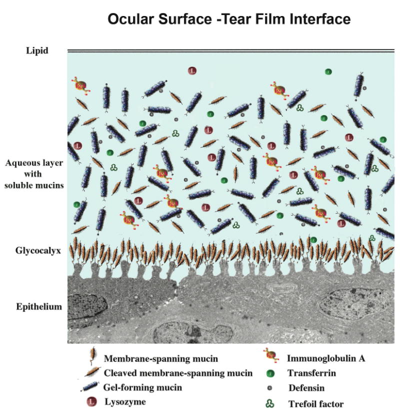
Diagram of the tear film and its interface with the ocular surface epithelium. The outermost lipid layer, the product of the meibomian gland, abuts the aqueous layer, with its soluble mucin components either secreted from conjunctival goblet cells, or shed from apical surfaces of corneal and conjunctival epithelial cells. A multitude of bactericidal and other proteins are present in the aqueous layer. The glycocalyx layer, which extends from the tips of surface ridges known as microplicae, is formed by membrane-associated mucins, which are tethered to the cells by a membrane-spanning domain and short cytoplasmic tail. The extracellular domains of these mucins are constituitively shed into the tear film.
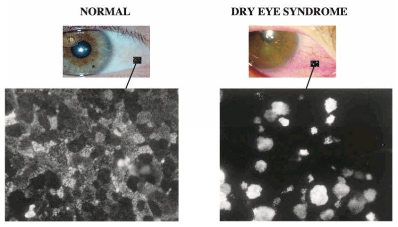
Impression cytology samples from normal subjects and dry eye patients (non-Sjögren's) showing change in localization pattern of binding of an antibody designated H185, which recognizes an O-acetylated sialic acid on the membrane-associated mucin MUC16. On the normal ocular surface, the antibody binds in a “patchwork” pattern, some cells binding the antibody intensely, others to a lesser degree, similar to the light, medium and dark cells seen by scanning electron microscopy. In dry eye patients, apical cell surface binding is lost and goblet cells bind the antibody intensely (see Danjo et al., 1998, for details).
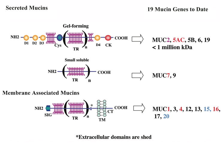
Diagram of the structure of the two classes of mucins—secreted and membrane associated. Nineteen mucins have been identified; those shown in red have been demonstrated to be expressed by the ocular surface epithelium by both in situ hybridization and immunohistochemistry. Those designated in blue have only been found in ocular surface epithelium by PCR and immunoblot analysis, and MUC2, in pink, has been found at low levels by PCR and immunoblot of tears. The characteristic common to both classes of mucins is the presence of tandem repeats (TR) of amino acids in their protein backbones, which may vary in number (n) between individuals (alleles are codominantly expressed). The tandem repeats are rich in serine and threonine, which are sites of O-glycosylation (YYY; in dark pink). Up to 80% of the mass of mucins can be O-glycans. Within the secreted category, two types of mucins are recognized—gel forming and small soluble. Gel-forming mucins are the largest proteins in the body; they have several D domains (D1, D2, etc.) and cysteine-rich domains (Cys, CK). The D domains allow for homomultimerization of individual gel-forming mucin molecules, which allows polymerization to form the viscous mucin gel. Membrane-associated mucins have a single membrane-spanning domain (TM) and a short cytoplasmic tail (CT). Their extracellular domains are constituitively shed into the tear film. (Figure after Argueso and Gipson, 2001.57)
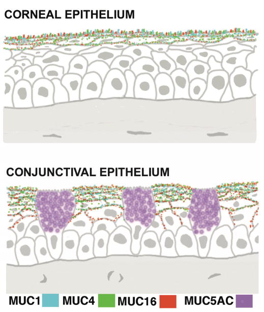
Diagram of sections of corneal and conjunctival epithelium showing the localization of expression of the membrane-spanning mucins MUC1, 4 and 16 in apical cells and the gel-forming mucin MUC5AC in goblet cells. There is very little expression of MUC4 in central epithelia. (Figure after Gipson, 2004.11)
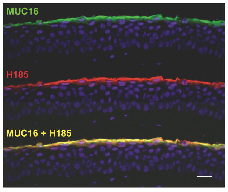
After 10 years of effort, H185 antibody was identified as a carbohydrate antigen on the membrane-associated mucin MUC16. The immunofluorescence micrographs of human corneal epithelium show colocalization of MUC16 and H185 antigen. This data and data showing co-immunoprecipitation of the same molecules with MUC16 and H185 antibodies demonstrate their identity. Bar = 25 μm.
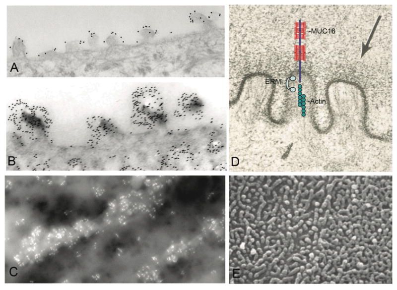
Electron micrographs of corneal epithelial surface microplicae. By transmission electron microscopy, MUC16 (A) and H185 (B) are localized by immunogold methods to the microplicae. Similar methods using field emission scanning electron microscopy were used to show that MUC16 is concentrated on the microplicae (C). Image in (D) summarizes a set of experiments in which we demonstrate that the cytoplasmic tail of MUC16 is tethered to the actin cytoskeleton through members of the actin linking family of proteins known as ERMs (ezrin, rodixin moesin, merlin). For reference, a lower magnification scanning electron microscopic view of surface microplicae is shown in (E).
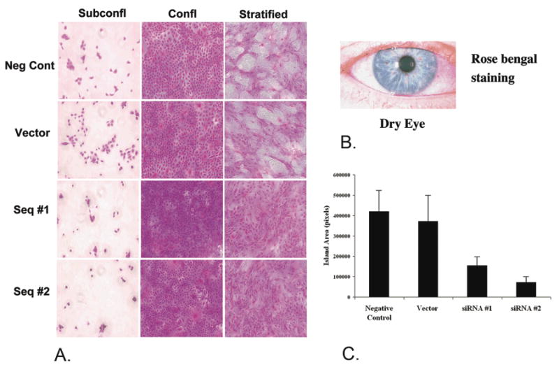
MUC16 provides a barrier to rose bengal dye as indicated by siRNA knockdown of the mucin in a human corneal limbal epithelial cell line. HCLE cells cultured to subconfluent or confluent stages do not express MUC16, and rose bengal dye penetrates all cells. After the cells are cultured seven additional days in serum continuing media, cells stratify and express MUC16. Islands of differentiated cells exclude the rose bengal dye. When MUC16 was knocked down by 80-90% as with Seq. #1 and Seq. #2, the islands that exclude the dye are diminished (A) as quantified in (C). For comparison, rose bengal stains areas of damaged ocular surface epithelium in dry eye (B). (For details of experiments see Blalock et al., In Press.48)
References
-
- Research in dry eye: Report of the Research Subcommittee of the International Dry Eye WorkShop in: Report of the International Dry Eye WorkShop (DEWS) Ocul Surf. 2007;5:179–193. - PubMed
-
- Williams K, Watsky M. Gap junctional communication in the human corneal endothelium and epithelium. Curr Eye Res. 2002;25:29–36. - PubMed
-
- Walcott B, Moore LC, Birzgalis A, et al. Role of gap junctions in fluid secretion of lacrimal glands. Am J Physiol Cell Physiol. 2002;282:C501–507. - PubMed
-
- Kinoshita S, Adachi W, Sotozono C, et al. Characteristics of the human ocular surface epithelium. Prog Retin Eye Res. 2001;20:639–673. - PubMed
-
- Paulsen FP, Schaudig U, Thale AB. Drainage of tears: impact on the ocular surface and lacrimal system. Ocul Surf. 2003;1:180–191. - PubMed
Publication types
MeSH terms
Substances
Grants and funding
LinkOut - more resources
Full Text Sources
Other Literature Sources
