. 2007 Oct;48(10):4407-20.
doi: 10.1167/iovs.07-0432.

SOD2 knockdown mouse model of early AMD

Affiliations

SOD2 knockdown mouse model of early AMD

Verline Justilien et al. Invest Ophthalmol Vis Sci. 2007 Oct.

Abstract

Purpose: To test the hypothesis that oxidative injury to the retinal pigment epithelium (RPE) may lead to retinal damage similar to that associated with the early stages of age-related macular degeneration (AMD).

Methods: A ribozyme that targets the protective enzyme manganese superoxide dismutase (MnSOD) was expressed in RPE-J cells, and adeno-associated virus (AAV) expressing the ribozyme gene was injected beneath the retinas of adult C57BL/6 mice. The RPE/choroid complex was examined for SOD2 protein levels and protein markers of oxidative damage using immunoblot analysis and LC MS/MS-identification of proteins and nitration sites. Lipids were extracted from retinal tissue and analyzed for the bis-retinoid compounds A2E and iso-A2E. The mice were analyzed by full-field electroretinography (ERG) for light response. Light and electron microscopy were used to measure cytological changes in the retinas.

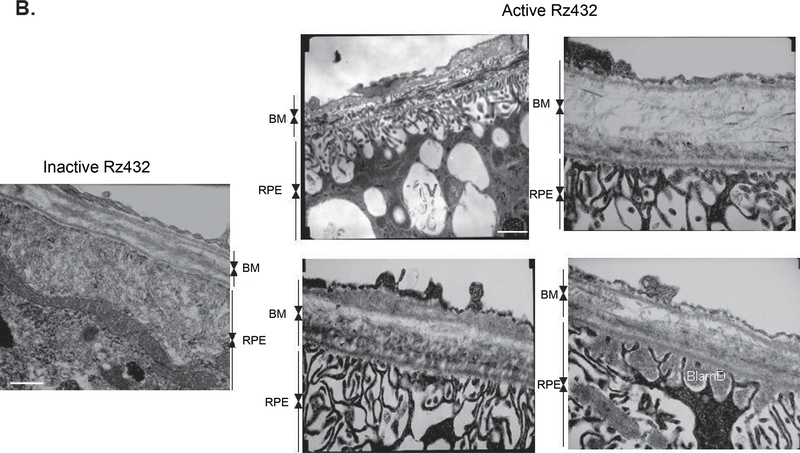
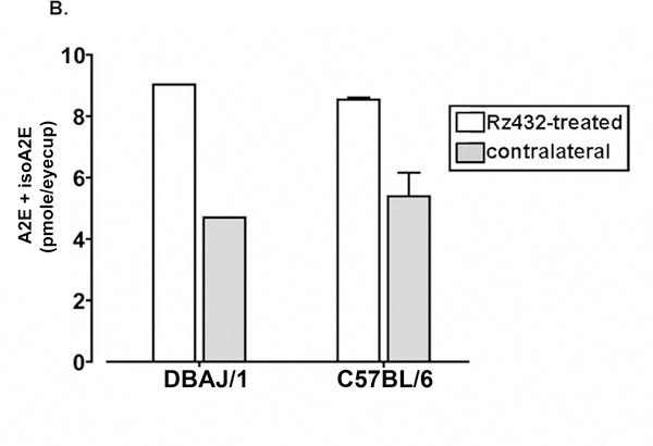
Results: The treatment of RPE-J cells with Rz432 resulted in decreased MnSOD mRNA and protein as well as increased levels of superoxide anion and apoptotic cell death. When delivered by AAV, Rz432 reduced MnSOD protein and increased markers of oxidative damage, including nitrated and carboxyethylpyrrole-modified proteins in the RPE-choroid of mice. Ribozyme delivery caused a progressive loss of electroretinograph response, vacuolization, degeneration of the RPE, thickening of Bruch's membrane, and shortening and disorganization of the photoreceptor outer and inner segments. Progressive thinning of the photoreceptor outer nuclear layer resulted from apoptotic cell death. Similar to the eyes of patients with AMD, ribozyme-treated eyes exhibited increased autofluorescence and elevated levels of A2E and iso-A2E, major bis-retinoid pigments of lipofuscin.

Conclusions: These results support the hypothesis that oxidative damage to the RPE may play a role in some of the key features of AMD.


Figures

Figure 1.
Figure 1.
Secondary structure of hammerhead ribozyme targeting the MnSOD and AAV packaging construct. (A) Sequence of MnSOD Rz432 and its MnSOD mRNA targeting sequence with cleavage site (thick arrow). Nucleotide position change to produce inactive MnSOD Rz432 (thin arrow). (B) The recombinant AAV cassette used to produce CBA-SOD2Rz432/CMV-GFP viral vector. TR, inverted terminal repeats; CMV, cytomegalovirus; CBA, chicken β-actin; SD/SA, splice donor/acceptor site; hGFP, humanized green fluorescent protein.
Figure 2.
Figure 2.
Effects of Rz432 on MnSOD expression in RPE-J cells. (A) Quantification of MnSOD transcript levels measured in triplicate with reverse transcription PCR. β-Actin amplified in the same reactions was used as an internal control. PCR products were quantitated by SYBR green staining. A significant decrease in MnSOD transcripts was observed by 24 hours after treatment with Rz432 (P < 0.05). (B) Western blot for MnSOD protein levels from Rz432 or GFP-treated cells. MnSOD mRNA and protein levels returned to normal after 4 days, because of the transient transfection. This experiment was performed twice, with essentially identical results.
Figure 3.
Figure 3.
Expression of Rz432 in RPE reduced MnSOD protein in the retina. (A) Localization of Rz432-GFP expression at 6 weeks after injection (green). Left: a whole-mounted retina viewed from the anterior surface. Inset: higher magnification showing GFP transduction of hexagonal RPE cells. Right: a transverse section through the retina counterstained with DAPI to show cell nuclei (blue). A single injection of the vector transduced almost the entire span of the RPE. Inset: higher magnification illustrates that cells of the RPE and not photoreceptors were transduced. (B) Rz432 expression reduces MnSOD protein levels in the RPE/choroid. Groups of mice were analyzed by Western blot for MnSOD levels at 6 weeks after injection (left). β-Actin was used as a loading control. Graph shows the ratio of MnSOD to β-actin signal (n = 6). Representative immunoblot of MnSOD protein from RPE/choroid of Rz432 and inactive Rz-treated eyes (right).
Figure 4.
Figure 4.
Suppression of MnSOD2 led to accumulation of markers of oxidative stress. Western blot analysis of nitrotyrosine (A) and carboxyethylpyrrole (B) were performed with protein extracts of posterior eye cups. Rz432-treated eyes show significantly increased staining of bands immunoreactive for nitrotyrosine and carboxyethylpyrrole (immunoblots). The nitrotyrosine and carboxyethylpyrrole bands were quantitated by scanning and measuring optical densities (scatterplots). (◇) Rz432 treated; (□) control. (P < 0.03 for nitrotyrosine and P < 0.045 for carboxyethylpyrrole; n = 6).
Figure 5.
Figure 5.
Treatment of wild-type mice with Rz432 led to reduced light response. (A) Scotopic full-field ERGs were measured at 1, 2, 4, and 6 months after injection in groups of adult wild-type C57BL/6 mice injected with AAV-Rz432 or AAV-GFP control vector. Representative ERG waveforms from the 1- and 6-month time points are shown. R (right eye), AAV-Rz432; L (left eye), AAV-GFP control vector. (B) Mice treated with Rz432 showed progressive loss of ERG response that was significant by 4 months after injection. The graph represents the ratio of the maximum a- and b-wave amplitudes of Rz432 to GFP control. *P < 0.05; **P < 0.005. Error bars, SEM.
Figure 6.
Figure 6.
Rz432 caused histologic damage to the outer retinas of mice. (A) Light micrographs of retinas of C57BL/6 mice at 1, 2, and 4 months after treatment with Rz432 or 4 months after treatment with GFP control vector. Rz432-treated retinas showed pigmentary changes (arrow-heads) and degeneration of the RPE (arrow) as well as progressive thinning of the photoreceptor nuclear layer. RPE, retinal pigmented epithelium; OS, photoreceptor outer segments; IS, photoreceptor inner segments; ONL, outer nuclear layer; INL, inner nuclear layer; IPL, inner plexiform layer. Scale bar, 50 μm. (B) Quantification of the thickness of the ONL. With the optic nerve head serving as a landmark, measurements were taken at 200-μm increments from the optic nerve on both the superior and inferior portions of the retina (n = 3 for each time point). Error bars, SEM *P < 0.05–P < 0.01. (C) Progressive loss of photoreceptor cells was due to apoptotic cell death. TUNEL staining was used to detect apoptotic nuclei in the retina at 6 weeks after Rz432 treatment (left). Green: Rz432-GFP expression; red: TUNEL-positive nuclei; blue, DAPI stained nuclei. (D) Quantification of apoptotic cell death by nucleosome release assay at 6 weeks after injection (right). *P < 0.003 (n = 6).
Figure 6.
Figure 6.
Rz432 caused histologic damage to the outer retinas of mice. (A) Light micrographs of retinas of C57BL/6 mice at 1, 2, and 4 months after treatment with Rz432 or 4 months after treatment with GFP control vector. Rz432-treated retinas showed pigmentary changes (arrow-heads) and degeneration of the RPE (arrow) as well as progressive thinning of the photoreceptor nuclear layer. RPE, retinal pigmented epithelium; OS, photoreceptor outer segments; IS, photoreceptor inner segments; ONL, outer nuclear layer; INL, inner nuclear layer; IPL, inner plexiform layer. Scale bar, 50 μm. (B) Quantification of the thickness of the ONL. With the optic nerve head serving as a landmark, measurements were taken at 200-μm increments from the optic nerve on both the superior and inferior portions of the retina (n = 3 for each time point). Error bars, SEM *P < 0.05–P < 0.01. (C) Progressive loss of photoreceptor cells was due to apoptotic cell death. TUNEL staining was used to detect apoptotic nuclei in the retina at 6 weeks after Rz432 treatment (left). Green: Rz432-GFP expression; red: TUNEL-positive nuclei; blue, DAPI stained nuclei. (D) Quantification of apoptotic cell death by nucleosome release assay at 6 weeks after injection (right). *P < 0.003 (n = 6).
Figure 7.
Figure 7.
Ultrastructural changes in the outer retina at 4 months after AAV-Rz432 treatment. (A) Active Rz432-treated retinas (A2, A4) compared with control inactive Rz432-injected retinas (A1, A3), the RPE appeared distended with increased vacuolization and irregularly shaped nuclei. The outer and inner segments of the photoreceptors were shortened and disorganized. (B) Electron micrographs of Rz432 treated retinas showing unusual deposits between the plasma and basement membrane of the RPE as well as disorganization and increased thickening in the layers of Bruch’s membrane. BlamD, basal laminar deposits. Scale bar (A1, A2) 10 μm; (A3, A4) 5 μm; (B) 0.5 μm.
Figure 7.
Figure 7.
Ultrastructural changes in the outer retina at 4 months after AAV-Rz432 treatment. (A) Active Rz432-treated retinas (A2, A4) compared with control inactive Rz432-injected retinas (A1, A3), the RPE appeared distended with increased vacuolization and irregularly shaped nuclei. The outer and inner segments of the photoreceptors were shortened and disorganized. (B) Electron micrographs of Rz432 treated retinas showing unusual deposits between the plasma and basement membrane of the RPE as well as disorganization and increased thickening in the layers of Bruch’s membrane. BlamD, basal laminar deposits. Scale bar (A1, A2) 10 μm; (A3, A4) 5 μm; (B) 0.5 μm.
Figure 8.
Figure 8.
Increased autofluorescence in oxidatively stressed retinas. (A) At 4.5 months after injection, flatmounts of the RPE/choroid/sclera were prepared from eyes fixed with paraformaldehyde and examined by fluorescence microscopy (excitation, 405 nm; emission, 590–650 nm) to detect autofluorescence in the presence of GFP fluorescence from the AAV vector. (B) Quantitation of A2E/iso-A2E in eyes of mice after ribozyme-mediated knockdown of SOD2. Mouse posterior eye cups were analyzed at age 4.5 months for DBAJ/1, which contains the Leu450 variant of RPE65, and at 5 to 6 months for C57BL/6J, which contains the Met450 variant of RPE65. Pigments were detected by HPLC with monitoring at 430 nm. A2E and iso-A2E levels were measured separately and summed. Results are based on single samples (4.5 months) or the mean (±SEM) of two samples (5–6 months). Each sample contained three to four eyes.
Figure 8.
Figure 8.
Increased autofluorescence in oxidatively stressed retinas. (A) At 4.5 months after injection, flatmounts of the RPE/choroid/sclera were prepared from eyes fixed with paraformaldehyde and examined by fluorescence microscopy (excitation, 405 nm; emission, 590–650 nm) to detect autofluorescence in the presence of GFP fluorescence from the AAV vector. (B) Quantitation of A2E/iso-A2E in eyes of mice after ribozyme-mediated knockdown of SOD2. Mouse posterior eye cups were analyzed at age 4.5 months for DBAJ/1, which contains the Leu450 variant of RPE65, and at 5 to 6 months for C57BL/6J, which contains the Met450 variant of RPE65. Pigments were detected by HPLC with monitoring at 430 nm. A2E and iso-A2E levels were measured separately and summed. Results are based on single samples (4.5 months) or the mean (±SEM) of two samples (5–6 months). Each sample contained three to four eyes.

Similar articles

Cited by

References

    1. Klein R, Klein BE, Jensen SC, Meuer SM. The five-year incidence and progression of age-related maculopathy: the Beaver Dam Eye Study. Ophthalmology. 1997;104:7–21. - PubMed
    1. Leibowitz HM, Krueger DE, Maunder LR, et al. The Framingham Eye Study monograph: an ophthalmological and epidemiological study of cataract, glaucoma, diabetic retinopathy, macular degeneration, and visual acuity in a general population of 2631 adults, 1973–1975. Surv Ophthalmol. 1980;24:335–610. - PubMed
    1. Mitchell P, Smith W, Attebo K, Wang JJ. Prevalence of age-relatedmaculopathy in Australia: the Blue Mountains Eye Study. Ophthalmology. 1995;102:1450–1460. - PubMed
    1. Vingerling JR, Dielemans I, Hofman A, et al. The prevalence ofage-related maculopathy in the Rotterdam Study. Ophthalmology. 1995;102:205–210. - PubMed
    1. Young RW. Pathophysiology of age-related macular degeneration. Surv Ophthalmol. 1987;31:291–306. - PubMed

Publication types

MeSH terms