. 2007 Oct;48(10):4664-70.
doi: 10.1167/iovs.07-0473.

Modulation of corneal epithelial innate immune response to pseudomonas infection by flagellin pretreatment

Affiliations

Modulation of corneal epithelial innate immune response to pseudomonas infection by flagellin pretreatment

Ashok Kumar et al. Invest Ophthalmol Vis Sci. 2007 Oct.

Abstract

Purpose: A prior study showed that Toll-like receptor (TLR)-5 recognizes Pseudomonas aeruginosa flagellin and triggers the production of proinflammatory cytokines in human corneal epithelial cells (HCECs). The present study was conducted to determine how the inflammatory response is modulated after TLR activation in HCECs.

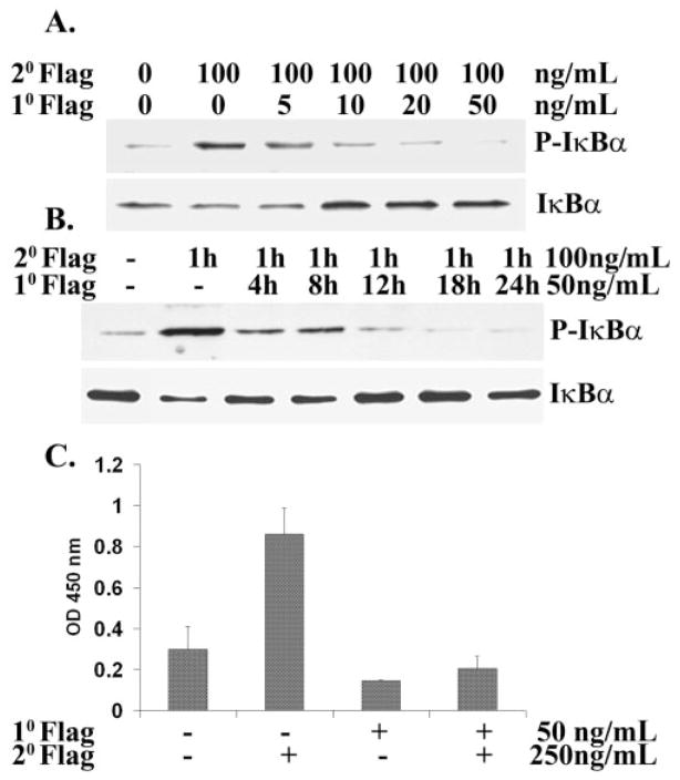
Methods: HUCL cells, a telomerase-immortalized HCEC line, and primary cultures of HCECs were pretreated with low-dose flagellin and then challenged, with either a high dose of flagellin or with Pseudomonas. NF-kappaB activation was determined by the extent of IkappaB-alpha phosphorylation and degradation and of nuclear p65 DNA binding. The amount of cytokines in the culture media was assessed by ELISA. The activation of p38 and JNK and the cellular expression of TLR5 were determined by Western blot analysis. Cell surface distribution of TLR5 was assessed by flow cytometry. The expression and secretion of antimicrobial peptides were assessed by semiquantitative RT-PCR and slot-blot analysis, respectively.

Results: Pre-exposure (12-24 hours) of HCECs to low-dose flagellin induced a state of tolerance, characterized by impaired activation of the NF-kappaB, p38, and JNK pathways and reduced production of IL-8 and TNF-alpha on subsequent challenge with a high dose of flagellin. Flagellin-induced tolerance did not alter the cellular level and surface distribution of TLR5. Furthermore, flagellin priming of HCECs dampened the inflammatory response of HCECs to live Pseudomonas. Pseudomonas-induced upregulation of antimicrobial genes such as hBD2 and LL-37 was augmented, even in tolerized HCECs.

Conclusions: Pre-exposure of the ocular surface to TLR agonists may induce protective mechanisms that not only modulate the host inflammatory response but also provide an innate defense against bacterial infection in the cornea.


Figures

Figure 1
Figure 1
Flagellin-elicited NF-κB activation in flagellin-pretreated HCECs: dose and time-dependent course studies. HUCL cells were incubated with various concentrations of flagellin for 24 hours: (A) with 50 ng/mL flagellin before a second 1-hour incubation with various concentrations of flagellin; (B) or with 50 ng/mL flagellin for various periods before a second 1-hour incubation with 100 ng/mL flagellin, with unstimulated cells as the control. (C) Total cell lysate was prepared after the second flagellin challenge and analyzed for phospho-IκB-α (IκB-α) and degradation (IκB-α) by immunoblot analysis. NF-κB p65 DNA-binding activity was detected by ELISA (n = 3) and colorimetry. Results are representative of those in three independent experiments. 1°, primary treatment; 2°, secondary treatment.
Figure 2
Figure 2
Flagellin-stimulated IκB-α phosphorylation and degradation and IL-8 secretion in HCECs. HUCL cells were stimulated with different concentrations of flagellin as indicated for 1 hour, and the cell lysates were subjected to SDS-PAGE, followed by phospho-IκBα (P-IκBα) and IκBα immunoblot analysis (A). IL-8 secretion from supernatant of cultured HUCL cells challenged with 10, 50, and 250 ng/mL flagellin for 12 hours was analyzed by ELISA (B). The amount of cytokines was normalized with protein concentration of cell lysate (ng/mg cell lysate). The data shown are representative of triplicate experiments.
Figure 3
Figure 3
Activation of MAPK signaling was inhibited in flagellin-tolerized cells. HUCL cells were either pretreated with 50 ng/mL flagellin or left untreated for 24 hours and then challenged with 250 ng/mL flagellin for the indicated times. Total cell lysates were blotted with antibodies specific for phospho-p38, -JNK, and -IκB-α. The cellular levels of these proteins were assessed in parallel with Western blot analysis. Flagellin-pretreated cells showed diminished MAPK signaling compared with untreated naive cells. The data shown are representative of duplicate experiments.
Figure 4
Figure 4
Expression of TLR5 in unstimulated and flagellin-tolerized HCECs. (A) HUCL cells were stimulated with flagellin (50 ng/mL) and, at the indicated times, were lysed for TLR5 detection by Western blot analysis. (B) HUCL cells were cultured with 50 ng/mL for 24 hours and were then stained with anti-TLR5 antibody or isotype control IgG and analyzed by flow cytometry. Cell surface expression of TLR5 was determined by flow cytometry. Control cells remained unstimulated. Data are representative of results in two independent experiments.
Figure 5
Figure 5
Flagellin-induced tolerance to a second flagellin or PA01 challenge in the production of TNF-α and IL-8. Primary HCECs (passage 3) or HUCL cells were cultured with or without 50 ng/mL flagellin for 24 hours. After being washed twice with PBS, the cells were stimulated with 250 ng/mL flagellin (A, C) or live PA01 (B, D) for 4 hours. TNF-α (A, B) and IL-8 (C, D) secretion into culture supernatants was assayed by ELISA. Data represent the mean ± SD of results in five independent experiments. *P < 0.001. 1°, primary treatment; 2°, secondary treatment.
Figure 6
Figure 6
Effect of flagellin pretreatment on PA01-mediated gene expression in HCECs. HUCL cells were cultured in KBM in the absence (Medium) or presence (Flag) of 50 ng/mL flagellin for 24 hours and then challenged with P. aeruginosa (multiplicity of infection, 100). At the indicated times, the cells were processed for semiquantitative RT-PCR, to assess mRNA expression of IL-8, TNF-α, hBD2, and LL-37, with GAPDH as the internal control (A). The secretion of antimicrobial peptides into the culture media was assessed by slot–blot analysis (B). Data are representative of results in two independent experiments.

References

    1. Jeng BH, McLeod SD. Microbial keratitis. Br J Ophthalmol. 2003;87:805–806. - PMC - PubMed
    1. Kurpakus-Wheater M, Kernacki KA, Hazlett LD. Maintaining corneal integrity how the “window” stays clear. Prog Histochem Cytochem. 2001;36:185–259. - PubMed
    1. Yu FS, Hazlett LD. Toll-like receptors and the eye. Invest Ophthalmol Vis Sci. 2006;47:1255–1263. - PMC - PubMed
    1. Medzhitov R, Preston-Hurlburt P, Janeway CA., Jr A human homologue of the Drosophila Toll protein signals activation of adaptive immunity (see comments) Nature. 1997;388:394–397. - PubMed
    1. Medzhitov R. Toll-like receptors and innate immunity. Nat Rev Immunol. 2001;1:135–145. - PubMed

Publication types

MeSH terms