

MRI patterns of atrophy associated with progression to AD in amnestic mild cognitive impairment
- PMID: 17898323
- PMCID: PMC2734138
- DOI: 10.1212/01.wnl.0000280575.77437.a2
MRI patterns of atrophy associated with progression to AD in amnestic mild cognitive impairment
Abstract
Objective: To compare the patterns of gray matter loss in subjects with amnestic mild cognitive impairment (aMCI) who progress to Alzheimer disease (AD) within a fixed clinical follow-up time vs those who remain stable.
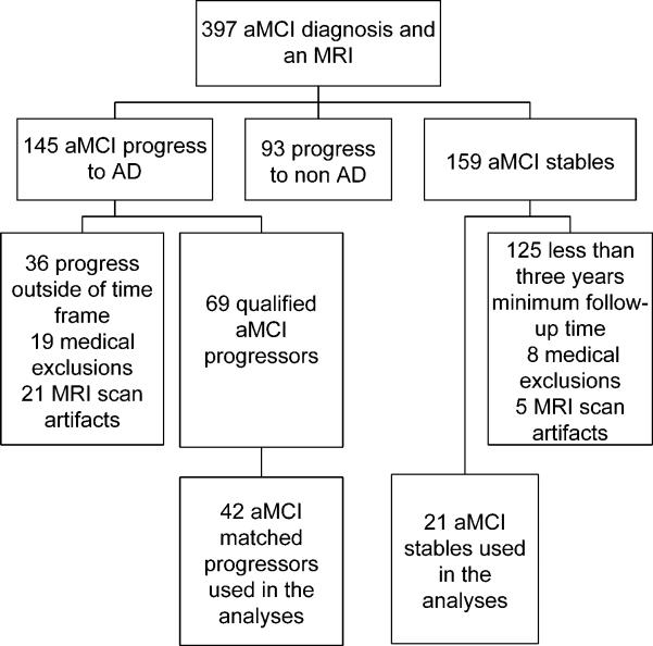
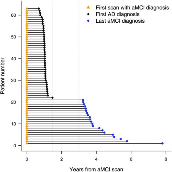
Methods: Twenty-one subjects with aMCI were identified from the Mayo Clinic Alzheimer's research program who remained clinically stable for their entire observed clinical course (aMCI-S), where the minimum required follow-up time from MRI to last follow-up assessment was 3 years. These subjects were age- and gender-matched to 42 aMCI subjects who progressed to AD within 18 months of the MRI (aMCI-P). Each subject was then age- and gender-matched to a control subject. Voxel-based morphometry (VBM) was used to assess patterns of gray matter atrophy in the aMCI-P and aMCI-S groups compared to the control group, and compared to each other.
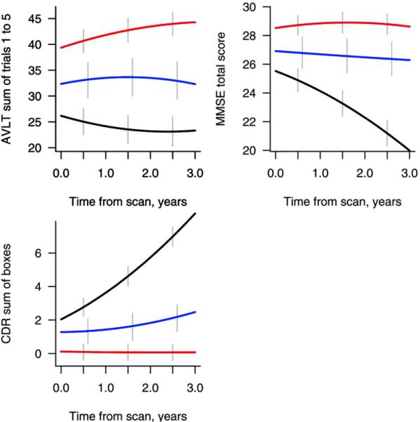
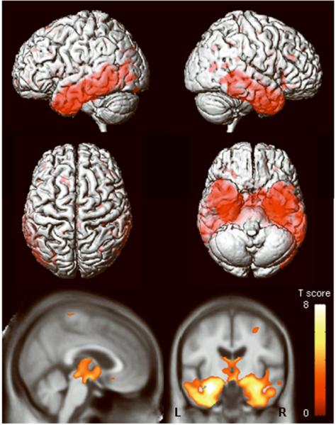
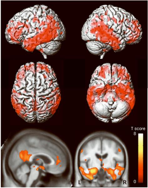
Results: The aMCI-P group showed bilateral loss affecting the medial and inferior temporal lobe, temporoparietal association neocortex, and frontal lobes, compared to controls. The aMCI-S group showed no regions of gray matter loss when compared to controls. When the aMCI-P and aMCI-S groups were compared directly, the aMCI-P group showed greater loss in the medial and inferior temporal lobes, the temporoparietal neocortex, posterior cingulate, precuneus, anterior cingulate, and frontal lobes than the aMCI-S group.
Conclusions: The regions of loss observed in subjects with amnestic mild cognitive impairment (aMCI) who progressed to Alzheimer disease (AD) within 18 months of the MRI are typical of subjects with AD. The lack of gray matter loss in subjects with aMCI who remained clinically stable for their entire observed clinical course is consistent with the notion that patterns of atrophy on MRI at baseline map well onto the subsequent clinical course.
Figures
Comment in
-
Is amnestic mild cognitive impairment always AD?Neurology. 2008 Feb 12;70(7):502-3. doi: 10.1212/01.wnl.0000299190.17488.b3. Neurology. 2008. PMID: 18268243 No abstract available.
References
-
- Petersen RC, Smith GE, Waring SC, Ivnik RJ, Tangalos EG, Kokmen E. Mild cognitive impairment: clinical characterization and outcome. Arch Neurol. 1999;56:303–308. - PubMed
-
- Petersen RC. Mild cognitive impairment as a diagnostic entity. J Intern Med. 2004;256:183–194. - PubMed
-
- Jicha GA, Parisi JE, Dickson DW, et al. Neuropathologic outcome of mild cognitive impairment following progression to clinical dementia. Arch Neurol. 2006;63:674–681. - PubMed
-
- Ganguli M, Dodge HH, Shen C, DeKosky ST. Mild cognitive impairment, amnestic type: an epidemiologic study. Neurology. 2004;63:115–121. - PubMed
Publication types
MeSH terms
Grants and funding
LinkOut - more resources
Full Text Sources
Other Literature Sources
Medical