. 2007 Oct 18;449(7164):928-32.
doi: 10.1038/nature06160. Epub 2007 Sep 26.

Arginine methylation at histone H3R2 controls deposition of H3K4 trimethylation

Affiliations

Arginine methylation at histone H3R2 controls deposition of H3K4 trimethylation

Antonis Kirmizis et al. Nature. .

Abstract

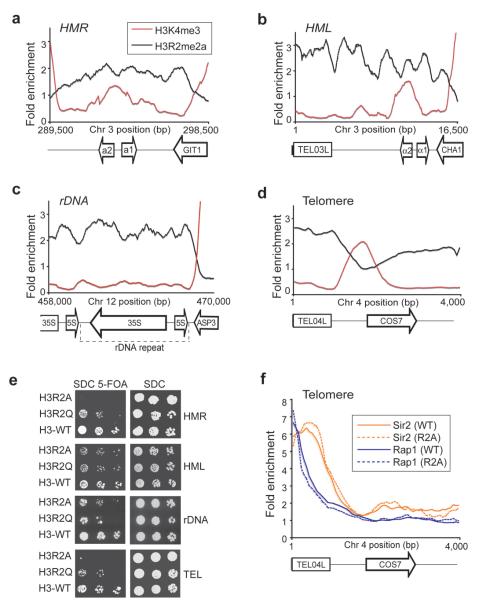
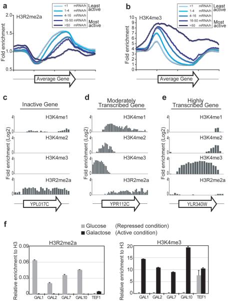
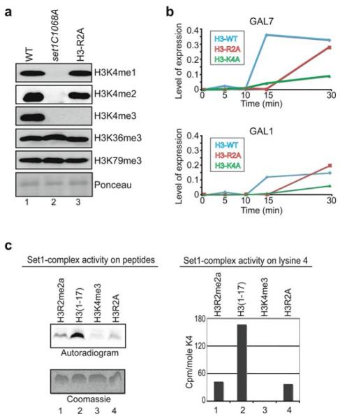
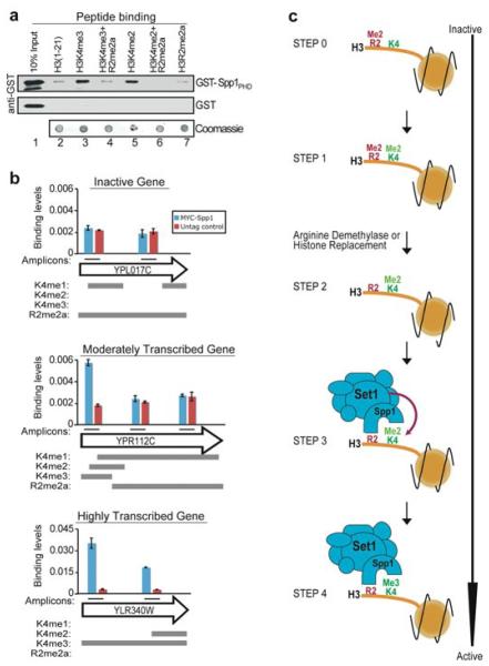
Modifications on histones control important biological processes through their effects on chromatin structure. Methylation at lysine 4 on histone H3 (H3K4) is found at the 5' end of active genes and contributes to transcriptional activation by recruiting chromatin-remodelling enzymes. An adjacent arginine residue (H3R2) is also known to be asymmetrically dimethylated (H3R2me2a) in mammalian cells, but its location within genes and its function in transcription are unknown. Here we show that H3R2 is also methylated in budding yeast (Saccharomyces cerevisiae), and by using an antibody specific for H3R2me2a in a chromatin immunoprecipitation-on-chip analysis we determine the distribution of this modification on the entire yeast genome. We find that H3R2me2a is enriched throughout all heterochromatic loci and inactive euchromatic genes and is present at the 3' end of moderately transcribed genes. In all cases the pattern of H3R2 methylation is mutually exclusive with the trimethyl form of H3K4 (H3K4me3). We show that methylation at H3R2 abrogates the trimethylation of H3K4 by the Set1 methyltransferase. The specific effect on H3K4me3 results from the occlusion of Spp1, a Set1 methyltransferase subunit necessary for trimethylation. Thus, the inability of Spp1 to recognize H3 methylated at R2 prevents Set1 from trimethylating H3K4. These results provide the first mechanistic insight into the function of arginine methylation on chromatin.


Figures

Figure 1
Figure 1. H3R2me2a associates with heterochromatin
(a-d) ChIP-on-chip analysis was performed in wild-type yeast cells (BY4741) grown to mid-log phase using antibodies to H3R2me2a and H3K4me3. The graphs show a moving average (window=15, step=1) of the H3R2me2a and H3K4me3 enrichment normalized to histone H3 occupancy at the right (HMR) and left mating type cassettes (HML), at the silent ribosomal DNA repeat region (rDNA) and at the left telomere of chromosome 4 (TEL04L). Values less than 1 represent regions that are not enriched. The arrows at the bottom of the graphs represent the location of genes and the direction they are transcribed. The rectangles at the bottom of the graphs correspond to the telomeric sequences. (e) Heterochromatin silencing assays were performed using cells from the H3R2A, H3R2Q, and isogenic wild-type (H3) yeast strains. Plates were photographed after 48 h incubation at 30 °C. (f) ChIP-on-chip analysis on telomeres using antibodies against Sir2p and Rap1p in WT and H3R2A strains. The graph shows a moving average (window=15, step=1) of the antibody enrichment as a ratio to Input at TEL04L.
Figure 2
Figure 2. H3R2me2a enrichment at euchromatic genes is mutually exclusive to H3K4me3
(a and b) ChIP-on-chip analysis was performed in wild-type yeast cells (BY4741) grown to mid-log phase using the anti-H3R2me2a and anti-H3K4me3 antibodies. The graphs represent composite profiles of H3R2me2a and H3K4me3 at 5065 genes, which were divided into five groups according to their transcriptional rate. (c-e) ChIP-on-chip analysis was performed as above using antibodies to H3K4me1, H3K4me2, H3K4me3, and H3R2me2a. The distribution of these modifications is compared at three differentially expressed genes. The name of each gene (arrow) is shown at the bottom of the graphs. (f) ChIP experiments were performed in yeast cells grown in either glucose (grey bars) or galactose as carbon source (black bars) using antibodies to H3R2me2a (left panel) and H3K4me3 (right panel). The precipitated DNA was analyzed by quantitative PCR using primers specific to the indicated genes. Standard errors were calculated for duplicate experiments. The induction of gene expression from glucose to galactose containing medium was monitored by RT-PCR analysis (see Supplementary Fig. S9).
Figure 3
Figure 3. H3R2me2a regulates the Set1-complex activity towards H3K4
(a) Whole yeast extracts prepared from the H3R2A (lane 3), set1C1068A (lane 2), and isogenic WT (lane 1) strains were analyzed by Western blotting using antibodies to H3K4me1, H3K4me2, H3K4me3, H3K36me3, or H3K79me3. Uniform loading was monitored by Ponceau stain. (b) Yeast cells expressing wild-type, R2A mutant, or K4A mutant histone H3 were grown to mid-log phase in media containing raffinose (inactive condition) and then shifted to media containing galactose (activating condition). RNA was prepared at the indicated times and analyzed by quantitative RT-PCR using primers specific to GAL7 (top panel) and GAL1 (bottom panel). Values show the expression of GAL7 or GAL1 normalized to the RNA levels of RTG2 whose expression remains unchanged. The experiment was repeated with similar results. (c) Yeast purified Set1-complex was incubated with 3H-SAM in the presence of an asymmetrically dimethylated H3R2 peptide (aa 1–17, lane 1), an unmodified peptide (aa 1-17, lane 2), a trimethylated H3K4 peptide (aa 1-17, lane 3), or an H3R2A mutant peptide (aa 1-17, lane 4). Peptides were analyzed for radioactive labelling using autoradiography (left panel) or sequential Edman degradation (right panel). Equal loading of peptides was monitored by Coomassie stain (left panel). The radioactivity released at lysine 4 of the H3K4me3 peptide (lane 3) was regarded as background signal and was subtracted from the lysine 4 signals of the other three peptides (right panel). The methylase assay was repeated three times with similar results.
Figure 4
Figure 4. H3R2me2a blocks the binding of Spp1 to methylated H3K4
(a) In vitro binding assays were performed using synthetic histone H3 N-terminal peptides (aa 1-21) and either recombinant GST-Spp1PHD (top panel) or GST alone (middle panel). The bound proteins were monitored by western analysis using a GST antibody. Peptide coupling was controlled by dot-blotting followed by Coomassie staining (bottom panel). (b) In vivo binding analysis of Spp1 was performed using ChIP assays followed by quantitative PCR. Chromatin from yeast cells expressing a tagged (Myc-Spp1) or an untagged (Control) form of Spp1 was immunoprecipitated with anti-Myc antibody. The analysed amplicons within each gene (arrow) are indicated by black lines. Standard errors were calculated for duplicate experiments. The grey bars below each plot show the distribution of mono-, di-, tri-H3K4 and H3R2me2a within each gene. (c) Model of how methylation at histone H3R2 controls trimethylation at H3K4.

References

    1. Kouzarides T. Chromatin modifications and their function. Cell. 2007;128:693–705. - PubMed
    1. Martin C, Zhang Y. The diverse functions of histone lysine methylation. Nat Rev Mol Cell Biol. 2005;6:838–49. - PubMed
    1. Wysocka J, Allis CD, Coonrod S. Histone arginine methylation and its dynamic regulation. Front Biosci. 2006;11:344–55. - PubMed
    1. Ruthenburg AJ, Allis CD, Wysocka J. Methylation of lysine 4 on histone H3: intricacy of writing and reading a single epigenetic mark. Mol Cell. 2007;25:15–30. - PubMed
    1. Sims RJ, 3rd, Reinberg D. Histone H3 Lys 4 methylation: caught in a bind? Genes Dev. 2006;20:2779–86. - PubMed

Publication types

MeSH terms

Substances

Associated data