. 2007 Dec;15(12):1422-31.
doi: 10.1007/s00167-007-0402-0. Epub 2007 Sep 27.

Description of the attachment geometry of the anteromedial and posterolateral bundles of the ACL from arthroscopic perspective for anatomical tunnel placement

Affiliations

Description of the attachment geometry of the anteromedial and posterolateral bundles of the ACL from arthroscopic perspective for anatomical tunnel placement

Joan W H Luites et al. Knee Surg Sports Traumatol Arthrosc. 2007 Dec.

Abstract

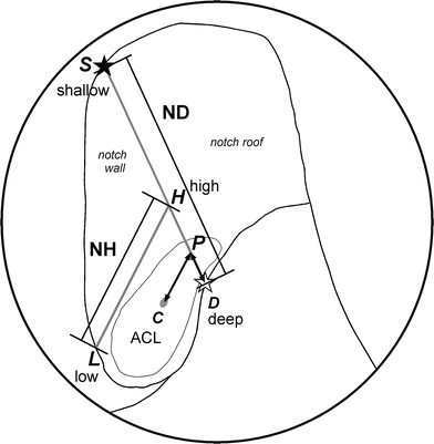
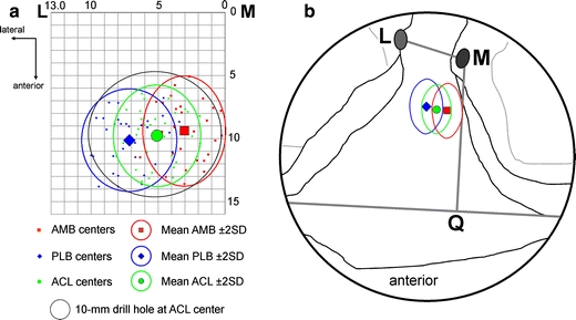
The anterior cruciate ligament (ACL) consists of an anteromedial bundle (AMB) and a posterolateral bundle (PLB). A reconstruction restoring the functional two-bundled nature should be able to approximate normal ACL function better than the most commonly used single-bundle reconstructions. Accurate tunnel positioning is important, but difficult. The purpose of this study was to provide a geometric description of the centre of the attachments relative to arthroscopically visible landmarks. The AMB and PLB attachment sites in 35 dissected cadaver knees were measured with a 3D system, as were anatomical landmarks of femur and tibia. At the femur, the mean ACL centre is positioned 7.9 +/- 1.4 mm (mean +/- 1 SD) shallow, along the notch roof, from the most lateral over-the-top position at the posterior edge of the intercondylar notch and from that point 4.0 +/- 1.3 mm from the notch roof, low on the surface of the lateral condyle wall. The mean AMB centre is at 7.2 +/- 1.8 and 1.4 +/- 1.7 mm, and the mean PLB centre at 8.8 +/- 1.6 and 6.7 +/- 2.0 mm. At the tibia, the mean ACL centre is positioned 5.1 +/- 1.7 mm lateral of the medial tibial spine and from that point 9.8 +/- 2.1 mm anterior. The mean AMB centre is at 3.0 +/- 1.6 and 9.4 +/- 2.2 mm, and the mean PLB centre at 7.2 +/- 1.8 and 10.1 +/- 2.1 mm. The ACL attachment geometry is well defined relative to arthroscopically visible landmarks with respect to the AMB and PLB. With simple guidelines for the surgeon, the attachments centres can be found during arthroscopic single-bundle or double-bundle reconstructions.


Figures

Fig. 1
Fig. 1
The orientation in the femoral notch, used in this study, is based on the recommendations of the ESKKA 1998 [4]. The notch depth (ND) is directed from shallow to deep in anatomic distal–proximal direction. The notch height (NH) is directed from low to high in anatomic dorsal–ventral direction. The notch wall is located at the medial side of the lateral condyle. The notch roof is the connection between the two condyles
Fig. 2
Fig. 2
a Distal view at a left femur. The high shallow and high deep points of the cartilage border were determined by placing the stylus at the point of an imaginary rectangular corner, indicating the separation between wall and roof, this corresponds with the 1.30 o’clock position in a left knee (10.30 o’clock in a right knee). b View at the medial side of a left lateral femoral condyle. The points that were digitized: AMB (white rounds) and PLB (black rounds) attachments; the cartilage border (grey asterisks), with the most distal, high shallow point (black asterisk) and most proximal, high deep point (white asterisk) indicating the separation between the notch wall on the lateral condyle and the notch roof
Fig. 3
Fig. 3
A schematic arthroscopic femoral view through the anteromedial portal. The femoral coordinate system is indicated. The first reference line, from the high deep point (D) to the high shallow point (S) on the cartilage edge separated the notch wall on the lateral condyle from the notch roof and defined the notch depth (ND). The second reference line, a perpendicular line, from the lowest point on the posterior cartilage edge (L) to the crossing point on the line DS (H) and defined the notch height (NH). The calculated attachment centre of the ACL (C) was projected on line DS (P). The absolute distances DP and CP were calculated, as were the distances relative to the reference lines (DP/DS and CP/LH). This was also done for the AMB and PLB, for the sake of clearness, only the centre of the entire ACL is depicted
Fig. 4
Fig. 4
Proximal view at the articular surfaces of the tibial condyles and the anterior intercondylar area. The points that were digitized: AMB (white rounds) and PLB (black rounds) attachments; the lateral tibial spine (light grey oval) and medial tibial spine (dark grey oval) and the most anterior points on the margin of the articular surface of the medial and lateral tibial condyles (white asterisks)
Fig. 5
Fig. 5
A schematic arthroscopic tibial view. The tibial coordinate system is indicated. The first reference line, from the medial spine (M) to the lateral spine (L), defined the interspinal distance (ID). The second reference line, from the medial spine (M) perpendicular to the crossing point (Q) at the anterior line between the most anterior points on the margin of the articular surface of the medial and lateral tibial condyles defined the anterior intercondylar length (AL). The calculated attachment centre (C) was projected on line ML (P). The absolute distances MP and CP were calculated, as were the distances relative to the reference lines (MP/ML and CP/MQ). This was also done for the AMB and PLB, for the sake of clearness, only the centre of the entire ACL is depicted
Fig. 6
Fig. 6
a A two-dimensional graph of the medial side of a right lateral femoral condyle with the individual centres, and the mean centres with the 95% Confidence Interval of the AMB (red squares), PLB (blue diamonds) and ACL (green dots). The position of a 12 mm drill hole at the ACL centre is also displayed. b A schematic arthroscopic view through the anteromedial portal with the mean centres and the 95% Confidence Interval areas of the AMB (red square with line), PLB (blue diamond with line) and ACL (green dot with line)
Fig. 7
Fig. 7
a A two-dimensional graph of a right anterior intercondylar tibial area with the individual centres, and the mean centres with the 95% Confidence Interval of the AMB (red squares), PLB (blue diamonds) and ACL (green dots). The position of a 10 mm drill hole at the ACL centre is also displayed. b A schematic arthroscopic view with the mean centres and the 95% Confidence Interval areas of the AMB (red square with line), PLB (blue diamond with line) and ACL (green dot with line)

Similar articles

Cited by

References

    1. {'text': '', 'ref_index': 1, 'ids': [{'type': 'DOI', 'value': '10.1007/s001670050041', 'is_inner': False, 'url': 'https://doi.org/10.1007/s001670050041'}, {'type': 'PubMed', 'value': '9335024', 'is_inner': True, 'url': 'https://pubmed.ncbi.nlm.nih.gov/9335024/'}]}
    2. Aglietti P, Buzzi R, Giron F, Simeone AJ, Zaccherotti G (1997) Arthroscopic-assisted anterior cruciate ligament reconstruction with the central third patellar tendon. A 5–8-year follow-up. Knee Surg Sports Traumatol Arthrosc 5(3):138–144 - PubMed
    1. {'text': '', 'ref_index': 1, 'ids': [{'type': 'DOI', 'value': '10.1007/BF01467913', 'is_inner': False, 'url': 'https://doi.org/10.1007/bf01467913'}, {'type': 'PubMed', 'value': '7584193', 'is_inner': True, 'url': 'https://pubmed.ncbi.nlm.nih.gov/7584193/'}]}
    2. Amis AA, Beynnon B, Blankevoort L, Chambat P, Christel P, Durselen L, Friederich N, Grood E, Hertel P, Jakob R (1994) Proceedings of the ESSKA Scientific Workshop on Reconstruction of the Anterior and Posterior Cruciate Ligaments. Knee Surg Sports Traumatol Arthrosc 2(3):124–132 - PubMed
    1. {'text': '', 'ref_index': 1, 'ids': [{'type': 'PubMed', 'value': '2005151', 'is_inner': True, 'url': 'https://pubmed.ncbi.nlm.nih.gov/2005151/'}]}
    2. Amis AA, Dawkins GP (1991) Functional anatomy of the anterior cruciate ligament. Fibre bundle actions related to ligament replacements and injuries. J Bone Joint Surg Br 73(2):260–267 - PubMed
    1. {'text': '', 'ref_index': 1, 'ids': [{'type': 'DOI', 'value': '10.1007/s001670050215', 'is_inner': False, 'url': 'https://doi.org/10.1007/s001670050215'}, {'type': 'PubMed', 'value': '9608456', 'is_inner': True, 'url': 'https://pubmed.ncbi.nlm.nih.gov/9608456/'}]}
    2. Amis AA, Jakob RP (1998) Anterior cruciate ligament graft positioning, tensioning and twisting. Knee Surg Sports Traumatol Arthrosc 6(Suppl 1):S2–S12 - PubMed
    1. {'text': '', 'ref_index': 1, 'ids': [{'type': 'PubMed', 'value': '6821989', 'is_inner': True, 'url': 'https://pubmed.ncbi.nlm.nih.gov/6821989/'}]}
    2. Arnoczky SP (1983) Anatomy of the anterior cruciate ligament. Clin Orthop 172:19–25 - PubMed

Publication types

LinkOut - more resources