

Biglycan and fibromodulin fragmentation correlates with temporal and spatial annular remodelling in experimentally injured ovine intervertebral discs
- PMID: 17899219
- PMCID: PMC2140141
- DOI: 10.1007/s00586-007-0497-5
Biglycan and fibromodulin fragmentation correlates with temporal and spatial annular remodelling in experimentally injured ovine intervertebral discs
Abstract
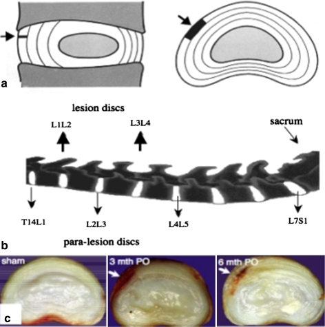
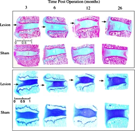
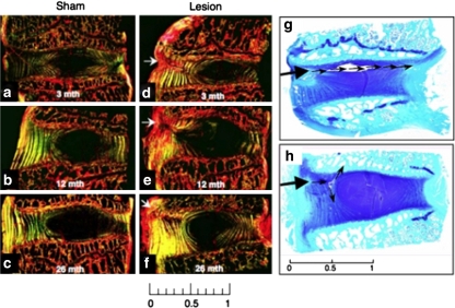
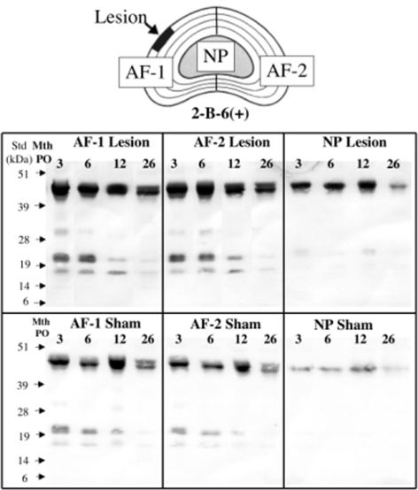
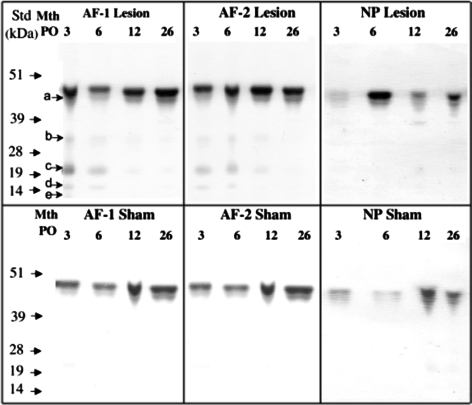
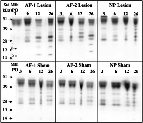
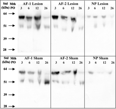
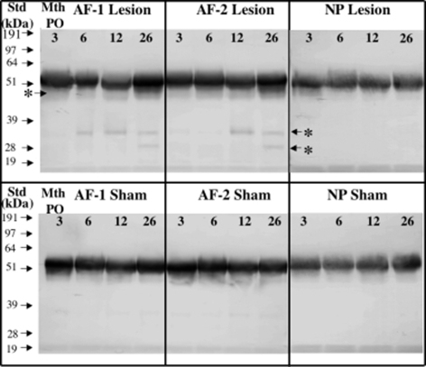
This study evaluated spatial and temporal extracellular matrix changes, induced by controlled surgical defects in the outer third of the annulus fibrosus (AF) of ovine intervertebral discs (IVDs). Thirty-two 4 year old sheep received a 4 mm deep x 10 mm wide standard annular surgical incision in the L1L2 and L3L4 IVDs (lesion group), 32 sheep were also subjected to the same surgical approach but the AF was not incised (sham-operated controls). Remodeling of the IVD matrix in the lesion and sham discs was assessed histochemically at 3, 6,12 and 26 month post operation (PO). Discs were also dissected into annular lesion site and contra-lateral AF and NP and equivalent zones in the sham sheep group, extracted with GuHCl, dialysed, freeze dried, digested with chondroitinase ABC/keratanase-I and aliquots examined for small leucine repeat proteoglycan (SLRP) core protein species by Western blotting using C-terminal antibodies to decorin, biglycan, lumican and fibromodulin and monoclonal antibody (Mab) 2B6 to unsaturated stub epitopes on chondroitin-4-sulphate generated by chondroitinase ABC. Masson Trichrome and Picrosirius red staining demonstrated re-organisation of the outermost collagenous lamellae in the incised discs 3-6 month PO. Toluidine blue staining also demonstrated a focal loss of anionic proteoglycan (PG) from the annular lesion 3-6 month PO with partial recovery of PG levels by 26 month. Specific fragments of biglycan and fibromodulin were associated with remodeling of the AF 12-26 month PO in the lesion IVDs but were absent from the NP of the lesion discs or all tissue zones in the sham animal group. Fragments of decorin were also observed in lesion zone extracts from 3 to 6 months but diminished after this. Isolation and characterization of the biglycan/fibromodulin fragments may identify them as prospective biomarkers of annular remodeling and characterization of the enzyme systems responsible for their generation may identify therapeutic target molecules.
Figures
Similar articles
-
A comparative analysis of the differential spatial and temporal distributions of the large (aggrecan, versican) and small (decorin, biglycan, fibromodulin) proteoglycans of the intervertebral disc.J Anat. 2001 Jan;198(Pt 1):3-15. doi: 10.1046/j.1469-7580.2001.19810003.x. J Anat. 2001. PMID: 11215765 Free PMC article.
-
A comparative evaluation of the small leucine-rich proteoglycans of pathological human intervertebral discs.Eur Spine J. 2012 May;21 Suppl 2(Suppl 2):S154-9. doi: 10.1007/s00586-012-2179-1. Epub 2012 Feb 23. Eur Spine J. 2012. PMID: 22358337 Free PMC article.
-
Age-related changes in the extracellular matrix of nucleus pulposus and anulus fibrosus of human intervertebral disc.Spine (Phila Pa 1976). 2009 Jan 1;34(1):10-6. doi: 10.1097/BRS.0b013e31818e5ddd. Spine (Phila Pa 1976). 2009. PMID: 19127156 Free PMC article.
-
Proteoglycans and diseases of soft tissues.Adv Exp Med Biol. 2014;802:49-58. doi: 10.1007/978-94-007-7893-1_4. Adv Exp Med Biol. 2014. PMID: 24443020 Review.
-
Molecular therapy of the intervertebral disc.Eur Spine J. 2006 Aug;15 Suppl 3(Suppl 3):S379-88. doi: 10.1007/s00586-006-0155-3. Epub 2006 Jul 12. Eur Spine J. 2006. PMID: 16835736 Free PMC article. Review.
Cited by
-
Extracellular matrix remodeling in experimental intervertebral disc degeneration.Acta Ortop Bras. 2013 May;21(3):144-9. doi: 10.1590/S1413-78522013000300003. Acta Ortop Bras. 2013. PMID: 24453658 Free PMC article.
-
Expression of lumican and fibromodulin following interleukin-1 beta stimulation of disc cells of the human temporomandibular joint.Eur J Histochem. 2011;55(2):e11. doi: 10.4081/ejh.2011.e11. Eur J Histochem. 2011. PMID: 22073367 Free PMC article. Clinical Trial.
-
Diverse and multifunctional roles for perlecan (HSPG2) in repair of the intervertebral disc.JOR Spine. 2024 Jul 29;7(3):e1362. doi: 10.1002/jsp2.1362. eCollection 2024 Sep. JOR Spine. 2024. PMID: 39081381 Free PMC article. Review.
-
Recent advances in annular pathobiology provide insights into rim-lesion mediated intervertebral disc degeneration and potential new approaches to annular repair strategies.Eur Spine J. 2008 Sep;17(9):1131-48. doi: 10.1007/s00586-008-0712-z. Epub 2008 Jun 27. Eur Spine J. 2008. PMID: 18584218 Free PMC article. Review.
-
Effects of initial boost with TGF-beta 1 and grade of intervertebral disc degeneration on 3D culture of human annulus fibrosus cells.J Orthop Surg Res. 2014 Aug 14;9:73. doi: 10.1186/s13018-014-0073-8. J Orthop Surg Res. 2014. PMID: 25116605 Free PMC article.
References
-
- {'text': '', 'ref_index': 1, 'ids': [{'type': 'DOI', 'value': '10.1093/glycob/cwf065', 'is_inner': False, 'url': 'https://doi.org/10.1093/glycob/cwf065'}, {'type': 'PubMed', 'value': '12213783', 'is_inner': True, 'url': 'https://pubmed.ncbi.nlm.nih.gov/12213783/'}]}
- Ameye L, Young MF (2002) Mice deficient in small leucine-rich proteoglycans: novel in vivo models for osteoporosis, osteoarthritis, Ehlers-Danlos syndrome, muscular dystrophy, and corneal diseases. Glycobiology 12(9):107R–116R - PubMed
-
- {'text': '', 'ref_index': 1, 'ids': [{'type': 'DOI', 'value': '10.1097/01.bor.0000240369.39713.af', 'is_inner': False, 'url': 'https://doi.org/10.1097/01.bor.0000240369.39713.af'}, {'type': 'PubMed', 'value': '16896297', 'is_inner': True, 'url': 'https://pubmed.ncbi.nlm.nih.gov/16896297/'}]}
- Ameye LG, Young MF (2006) Animal models of osteoarthritis: lessons learned while seeking the “Holy Grail”. Curr Opin Rheumatol 18(5):537–547 - PubMed
-
- Bancroft JD, Cook HC (eds) (1994) Manual of histological techniques and their diagnostic application, Chapt. 3. Churchill-Livingstone, London, p 42
-
- {'text': '', 'ref_index': 1, 'ids': [{'type': 'DOI', 'value': '10.1097/00007632-200212010-00002', 'is_inner': False, 'url': 'https://doi.org/10.1097/00007632-200212010-00002'}, {'type': 'PubMed', 'value': '12461389', 'is_inner': True, 'url': 'https://pubmed.ncbi.nlm.nih.gov/12461389/'}]}
- Boos N, Weissbach S, Rohrbach H, Weiler C, Spratt KF, Nerlich AG (2002) Classification of age-related changes in lumbar intervertebral discs: 2002 Volvo award in basic science. Spine 27(23):2631–2644 - PubMed
-
- {'text': '', 'ref_index': 1, 'ids': [{'type': 'PubMed', 'value': '7660243', 'is_inner': True, 'url': 'https://pubmed.ncbi.nlm.nih.gov/7660243/'}]}
- Buckwalter JA (1995) Aging and degeneration of the human intervertebral disc. Spine 20(11):1307–1314 - PubMed
Publication types
MeSH terms
Substances
LinkOut - more resources
Full Text Sources
Medical
Miscellaneous
