

doi: 10.1021/np070088v.
Epub 2007 Sep 28.
Induction of apoptosis by diterpenes from the soft coral Xenia elongata
Affiliations
- PMID: 17900165
- PMCID: PMC2866001
- DOI: 10.1021/np070088v
Item in Clipboard
Induction of apoptosis by diterpenes from the soft coral Xenia elongata
J Nat Prod.
2007 Oct.
Abstract
Four new diterpenes ( 1- 4) were isolated from the soft coral Xenia elongata using a novel cell-based screen for apoptosis-inducing, potential anticancer compounds. The molecular structures of the diterpenes were determined using a combination of NMR and mass spectrometry. The bioactivities were confirmed using a specific apoptosis induction assay based on genetically engineered mammalian lines with differential, defined capacities for apoptosis. The diterpenes induce apoptosis in micromolar concentrations. This is the first report of apoptosis induction by marine diterpenes in xenicane skeletons.
Figures
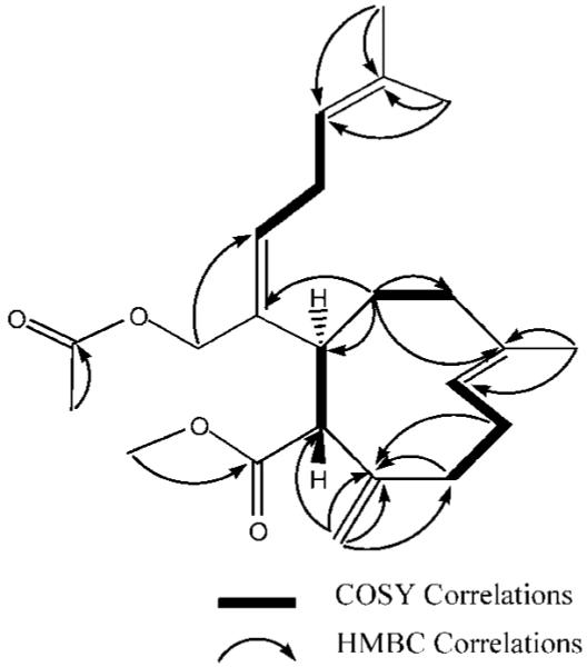
Key HMBC and selected COSY correlations for compound 2.
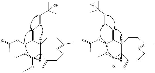
Selected ROESY cross-peaks for compounds 3 and 4.
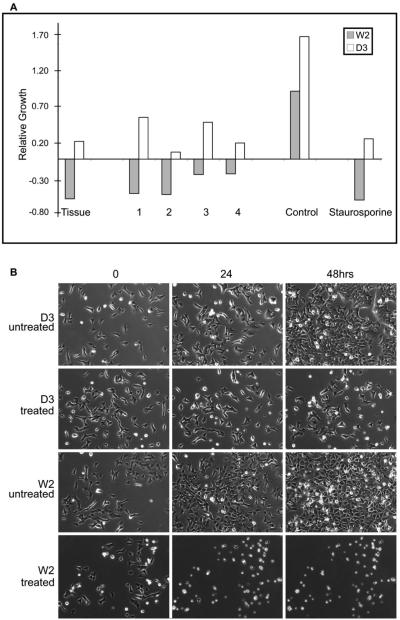
(A) Change in relative W2 and D3 cell viability by MTT assay 48 h after addition of whole tissue extract, 1, 2, 3, and 4 (1 12, 2 29, 3 12, 4 11 μM). Staurosporine (0.1 μM), a known apoptosis inducer, and untreated cells were used as positive and negative controls. Values for the compounds are an average of five wells, with a standard deviation of less than 15% of the mean. (B) Compound 1 is a specific inducer of apoptosis in mammalian cells. The apoptosis-competent W2 cells and the apoptosis -resistant D3 cells were cultured in the presence or absence of compound 1 (12 μM) under normal physiological conditions (DMEM with 10% FBS, 5% CO2 at 37 °C), and the cell viability was monitored by time-lapse microscopy. D3 cells were viable in the presence of compound 1 beyond 48 h, whereas the apoptosis-competent W2 cells showed massive apoptosis induction and significant loss of viability by 24 h of treatment.
References
-
- Andrianasolo EH, France D, Cornell-Kennon S, Gerwick WH. J. Nat. Prod. 2006;69:576–579. - PubMed
-
- Crews P, Gerwick W, Schmitz F, France D, Bair K, Wright A, Hallock Y. Pharm. Biol. 2005;41(Suppl. 1):39–52.
-
- Adams J. Genes Dev. 2003;17:2418–2495. - PubMed
-
- Danial N, Korsmeyer S. Cell. 2004;116:205–219. - PubMed
-
- Gelinas C, White E. Genes Dev. 2005;19:1263–1268. - PubMed
Publication types
MeSH terms
Substances
Grants and funding
LinkOut - more resources
Full Text Sources
Other Literature Sources
