. 2007 Dec;150(3):407-15.
doi: 10.1111/j.1365-2249.2007.03511.x. Epub 2007 Sep 27.

A role for T cell-derived interleukin 22 in psoriatic skin inflammation

Affiliations

A role for T cell-derived interleukin 22 in psoriatic skin inflammation

K Boniface et al. Clin Exp Immunol. 2007 Dec.

Abstract

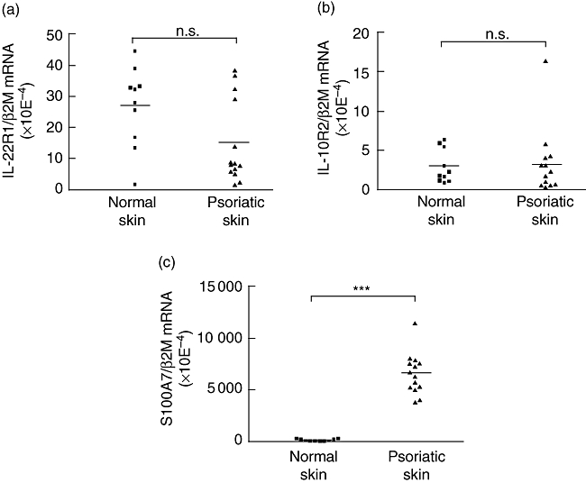
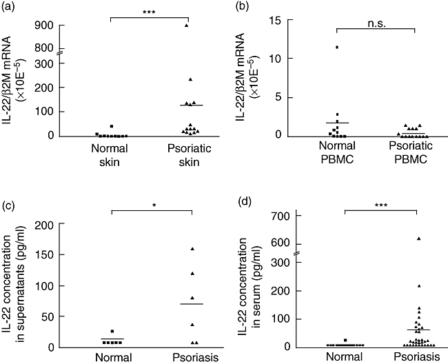
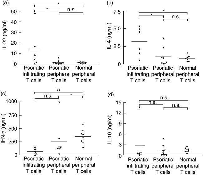
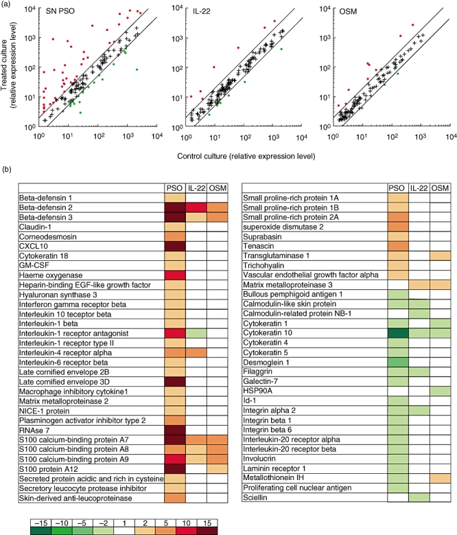
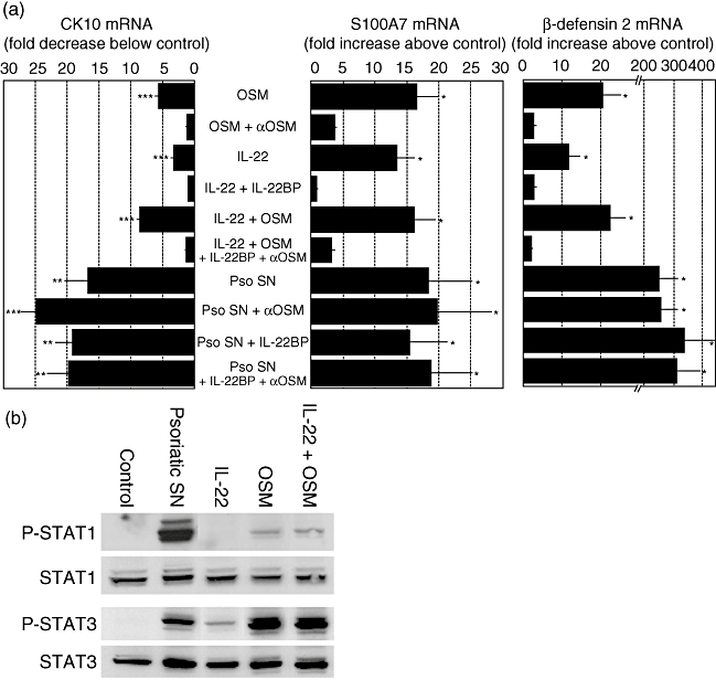
Interleukin (IL)-22 is a T cell-derived cytokine that has been reported recently to induce cutaneous inflammation in an experimental murine model of psoriasis, and to induce in vitro an inflammatory-like phenotype. In the present study, we assessed the presence of IL-22 and the IL-22 receptor 1 (IL-22R1) in skin lesions, skin-derived T cells, as well as IL-22 levels in sera from patients with psoriasis. IL-22R1 and IL-10R2 transcripts are expressed at a similar level in psoriatic and healthy skin. In contrast, IL-22 mRNA expression was up-regulated in psoriatic skin lesions compared to normal skin, whereas IL-22 mRNA levels in peripheral blood mononuclear cells from psoriatic patients and normal subjects were similar. Circulating IL-22 levels were significantly higher in psoriatic patients than in normal subjects. T cells isolated from psoriatic skin produced higher levels of IL-22 in comparison to peripheral T cells isolated from the same patients. IL-10 was expressed at similar levels in skin biopsies and peripheral blood mononuclear cells of psoriatic patients and normal subjects. Finally, we show here that supernatants of lesional psoriatic skin-infiltrating T cells induce an inflammatory response by normal human epidermal keratinocytes, resembling that observed in psoriatic lesions. Taken together, the results reported in this study indicate that IL-22 is a cytokine produced by skin-infiltrating lymphocytes that is potentially involved in initiation and/or maintenance of the pathogenesis of psoriasis.


Figures

Fig. 1
Fig. 1
Expression of the interleukin (IL)-22 receptor subunits in normal or lesional psoriatic skin. Total RNA was extracted from normal and psoriatic skin, reverse-transcribed, and IL-22R1 (a), IL-10R2 (b) and S100A7 (c) mRNA relative expressions were quantified by real-time reverse transcription–polymerase chain reaction. β-2 microglobulin was used as a housekeeping gene to normalize gene expression. ***P < 0·001, n.s.: not significant, based on a Mann–Whitney test.
Fig. 2
Fig. 2
Interleukin (IL)-22 mRNA and protein detection in skin biopsies, peripheral blood mononuclear cells (PBMC) and sera of normal or psoriatic patients. Total RNA was extracted from skin biopsies (a) or PBMC (b) from normal and psoriatic patients and reverse-transcribed. IL-22 mRNA relative expression was quantified by real-time reverse transcription–polymerase chain reaction. β-2 microglobulin was used as a housekeeping gene to normalize gene expression. ***P < 0·001, n.s.: not significant, based on a Mann–Whitney test. (c) Biopsies from normal skin or lesional psoriatic skin were cultured for 40 h and IL-22 concentration was measured in culture supernatants as in (a). *P < 0·05 based on a Mann–Whitney test. (d) IL-22 concentration was determined in the sera of healthy and psoriatic patients by enzyme-linked immunosorbent assay. ***P < 0·001 based on a Mann–Whitney test.
Fig. 3
Fig. 3
Production of interleukin (IL)-22, IL-4, interferon (IFN)-γ and IL-10 by lesional skin infiltrating T lymphocytes. T cells infiltrating lesional psoriatic skin and peripheral T cells of control or psoriatic patients were expanded for 10–14 days in the presence of Expander beads® and 10 ng/ml IL-2, and then activated for 24 h with IL-2, anti-CD3 and anti-CD28 monoclonal antibodies. IL-22 (a), IL-4 (b), IFN-γ (c) and IL-10 (d) concentrations were measured in culture supernatants by enzyme-linked immunosorbent assay. *P < 0·05, **P < 0·01, n.s.: not significant, based on a Mann–Whitney test.
Fig. 4
Fig. 4
Proinflammatory activities of psoriatic skin infiltrating T cells supernatants on normal human epidermal keratinocytes (NHEK). NHEK were cultured for 24 h with or without 20% supernatants of activated psoriatic skin infiltrating T cells (SN PSO) or 10 ng/ml of IL-22 or oncostatin M (OSM). The expression of 154 genes of potential interest for skin physiology was compared using home-made cDNA microarrays [27]. (a) The relative expression level in treated culture versus control culture is plotted. (b) List of regulated genes. The induced modulation was expressed as the ratio of the signal intensities for treated cells over unstimulated cells. The induced genes are shown in red, the suppressed ones in green. Representative results from one of three independent experiments, performed with three different supernatants of activated psoriatic skin infiltrating T cells (PSD), are shown.
Fig. 5
Fig. 5
Modulation of S100A7, β-defensin 2 and cytokeratin 10 (CK10) mRNA expression by psoriatic skin infiltrating T cells supernatants. (a) Normal human epidermal keratinocytes (NHEK) were cultured for 24 h with or without 20% supernatants of activated psoriatic skin infiltrating T cells (Pso SN), 10 ng/ml of interleukin (IL)-22, 10 ng/ml of oncostatin M (OSM), 1·5 μg/ml of IL-22BP or 40 μg/ml of anti-OSM blocking monoclonal antibody. Total RNA was extracted and reverse-transcribed. S100A7, β-defensin 2 and cytokeratin 10 mRNA relative expressions were quantified by real-time reverse transcription–polymerase chain reaction. β-2 microglobulin was used as a housekeeping gene to normalize gene expression. Data are mean ± standard error of the mean of three independent experiments. *P < 0·05; **P < 0·01; ***P < 0·001, based on a one-way analysis of variance and Newman–Keuls test. (b) NHEK were cultured 30 min with or without 20% supernatants of activated psoriatic skin infiltrating T cells or 10 ng/ml of IL-22 and/or OSM. Phospho-signal transducer and activator of transcription 1 (P-STAT1), P-STAT3, STAT1 and STAT3 protein levels were determined by Western blotting. Representative results from one of three independent experiments are shown.

References

    1. Lew W, Bowcock AM, Krueger JG. Psoriasis vulgaris: cutaneous lymphoid tissue supports T-cell activation and ‘Type 1’ inflammatory gene expression. Trends Immunol. 2004;25:295–305. - PubMed
    1. Gearing AJ, Fincham NJ, Bird CR, et al. Cytokines in skin lesions of psoriasis. Cytokine. 1990;2:68–75. - PubMed
    1. Boyman O, Conrad C, Tonel G, Gilliet M, Nestle FO. The pathogenic role of tissue-resident immune cells in psoriasis. Trends Immunol. 2007;28:51–7. - PubMed
    1. Boniface K, Diveu C, Morel F, et al. Oncostatin M secreted by skin infiltrating T lymphocytes is a potent keratinocyte activator involved in skin inflammation. J Immunol. 2007;178:4615–22. - PubMed
    1. Schlaak JF, Buslau M, Jochum W, et al. T cells involved in psoriasis vulgaris belong to the Th1 subset. J Invest Dermatol. 1994;102:145–9. - PubMed

Publication types

MeSH terms