. 2007 Sep 27:3:27.
doi: 10.1186/1744-8069-3-27.

Transformation of the output of spinal lamina I neurons after nerve injury and microglia stimulation underlying neuropathic pain

Affiliations

Transformation of the output of spinal lamina I neurons after nerve injury and microglia stimulation underlying neuropathic pain

A Florence Keller et al. Mol Pain. .

Abstract

Background: Disinhibition of neurons in the superficial spinal dorsal horn, via microglia - neuron signaling leading to disruption of chloride homeostasis, is a potential cellular substrate for neuropathic pain. But, a central unresolved question is whether this disinhibition can transform the activity and responses of spinal nociceptive output neurons to account for the symptoms of neuropathic pain.

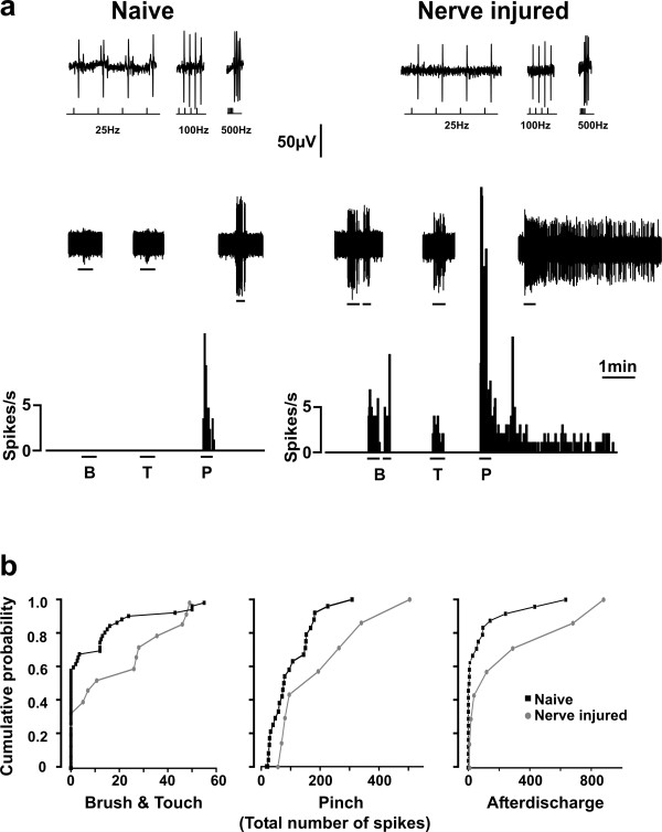
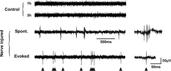
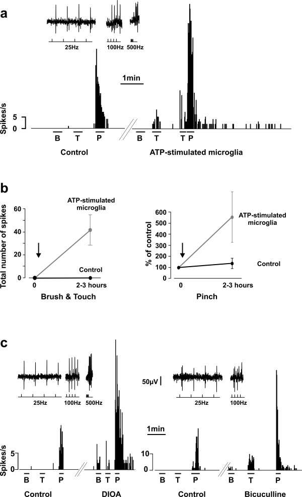
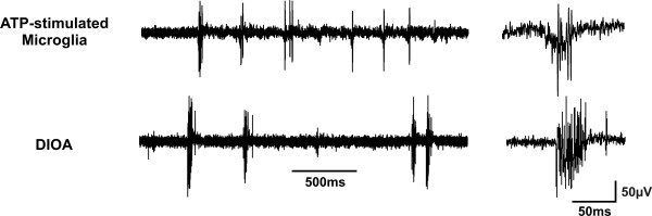
Results: Here we show that peripheral nerve injury, local spinal administration of ATP-stimulated microglia or pharmacological disruption of chloride transport change the phenotype of spinal lamina I output neurons, causing them to 1) increase the gain of nociceptive responsiveness, 2) relay innocuous mechanical input and 3) generate spontaneous bursts of activity. The changes in the electrophysiological phenotype of lamina I neurons may account for three principal components of neuropathic pain: hyperalgesia, mechanical allodynia and spontaneous pain, respectively.

Conclusion: The transformation of discharge activity and sensory specificity provides an aberrant signal in a primarily nociceptive ascending pathway that may serve as a basis for the symptoms of neuropathic pain.


Figures

Figure 1
Figure 1
Experimental paradigm. a. Schematic representation of the experimental setting to record from single antidromically identified lamina I projection neurons. Cells were recorded from control animals and animals that received a chronic constriction injury of the sciatic nerve. b. Confirmation of recording from lamina I projection neuron. Top: extracellular single unit recordings from a lamina I neuron showing one-for-one following of a train of antidromic stimuli (lower traces mark the stimulus; 1 mA, 200 μs duration; up to 500 Hz) delivered from the electrode positioned in the lateral parabrachial nucleus. Conduction distance was 100 mm. bottom: collision of the first of 4 antidromic action potentials (25 Hz) with an orthodromic action potential (*) occurring within the critical interval. The arrow points to the position where the first antidromic action potential would have occurred in absence of the orthodromic action potential (as in the trace on the left). c. Graph showing results of nociceptive reflexes to mechanical stimuli of the rats included in the current study. Peripheral nerve injury (N = 12) caused a significant reduction of the withdrawal threshold to mechanical stimulation of the hind paw. Nerve injured animals were taken between 16 and 24 days post-injury. Animals were anesthetized with pentobarbital or ketamine/xylazine and single unit extracellular recording was performed from lamina I projection neurons identified by antidromic stimulation from the parabrachial nucleus.
Figure 2
Figure 2
Nerve injury alters the sensory specificity of lamina I projection neurons. a. The majority (79%) of lamina I projection neurons in naïve rats were nociceptive specific whereas the majority (58%) of those recorded in nerve injured rats responded to both noxious and innocuous stimuli. The rate meter records show representative responses to natural stimuli (B = Brush; T = Touch; P = Pinch) of two identified lamina I projection neurons in a naïve rat and a rat with nerve injury. The inset shows the responses of the cells to trains of stimuli delivered in the parabrachial nucleus at decreasing interspike intervals; the cells followed > 500 Hz stimulation confirming antidromic activation (see methods and Fig. 1b). b. The cumulative probability plots show a significant increase in the response to innocuous (data from responses to brush and touch were pooled) and noxious (pinch) stimulation of the hind paw in lumbar spinal lamina I projection neurons after nerve injury. Results are expressed as total number of spikes during the stimulus for response to Brush/Touch and Pinch and throughout the duration of the afterdischarge (in other words, area under the curve).
Figure 3
Figure 3
Occurrence of spontaneous bursts of activity in lamina I projection neurons following nerve injury. Continuous records showing an example of an epoch with spontaneous bursts of activity occurring in lamina I projection neurons after nerve injury (6 out of 8 where ongoing activity was measured over a sufficiently long period to obtain a quantitative measure). The bursts were characterized by spikes arising on top of a slower wave, which contrasted with the evoked activity in response to touch (lower trace; arrowheads indicated touch stimuli). Such bursts were virtually absent in control animals even after several hours of recording. The insets on the right show examples of spontaneous and evoked activity on a faster time scale.
Figure 4
Figure 4
Stimulated microglia, disruption of chloride homeostasis and bicuculline alter the sensory selectivity of nociceptive specific lamina I projection neurons. a. Rate meter records at the top show the response of an identified lamina I projection neuron to natural mechanical stimulation of the receptive field (B = Brush; T = Touch; P = Pinch). The inset shows antidromic spikes from the parabrachial nucleus following our protocol for identification of projection neurons (see methods and Fig. 1b). b. Graphs showing the population data (values indicate mean ± SEM). To avoid biasing the results because of heterogeneity in responses between cells, values are expressed as a percent of control response for each cell. While none of 30 nociceptive specific lamina I projection neuron tested showed occurrence of responses to innocuous input (neither brush or touch) in control conditions after up to 4 hours of recording, all four nociceptive specific cells tested showed a significant response to innocuous input (brush and touch) within 2–3 h of local administration of ATP-stimulated microglia on the surface of the lumbar spinal cord. c. Blockade of cation-chloride co-transporters with local spinal administration of DIOA, or blockade of GABAA receptors with bicuculline unmasked innocuous input (brush and touch) to nociceptive specific lamina I projection neurons in control animals.
Figure 5
Figure 5
Stimulated microglia or disruption of chloride homeostasis provoke the occurrence of spontaneous bursts of activity in lamina I projection neurons. Continuous records showing the appearance of spontaneous bursts of activity in lamina I projection neurons after local administration of ATP-stimulated microglia or DIOA. The bursts were comparable to those observed in lamina I neurons from nerve-injured rats (see Fig. 3). Such burst were virtually absent in control conditions even after several hours of recording. The insets on the right show example bursts on a faster time scale.

References

    1. Baron R. Mechanisms of disease: neuropathic pain--a clinical perspective. Nat Clin Pract Neurol. 2006;2:95–106. doi: 10.1038/ncpneuro0113. - DOI - PubMed
    1. Backonja MM, Stacey B. Neuropathic pain symptoms relative to overall pain rating. J Pain. 2004;5:491–497. doi: 10.1016/j.jpain.2004.09.001. - DOI - PubMed
    1. Campbell JN, Meyer RA. Mechanisms of neuropathic pain. Neuron. 2006;52:77–92. doi: 10.1016/j.neuron.2006.09.021. - DOI - PMC - PubMed
    1. Woolf CJ, Salter MW. Neuronal plasticity: increasing the gain in pain. Science. 2000;288:1765–1769. doi: 10.1126/science.288.5472.1765. - DOI - PubMed
    1. Moore KA, Kohno T, Karchewski LA, Scholz J, Baba H, Woolf CJ. Partial peripheral nerve injury promotes a selective loss of GABAergic inhibition in the superficial dorsal horn of the spinal cord. J Neurosci. 2002;22:6724–6731. - PMC - PubMed

Publication types

MeSH terms

LinkOut - more resources