. 2007 Sep 27:7:178.
doi: 10.1186/1471-2148-7-178.

Molecular evolution of Phox-related regulatory subunits for NADPH oxidase enzymes

Affiliations

Molecular evolution of Phox-related regulatory subunits for NADPH oxidase enzymes

Tsukasa Kawahara et al. BMC Evol Biol. .

Abstract

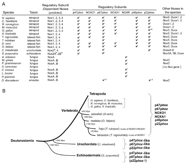
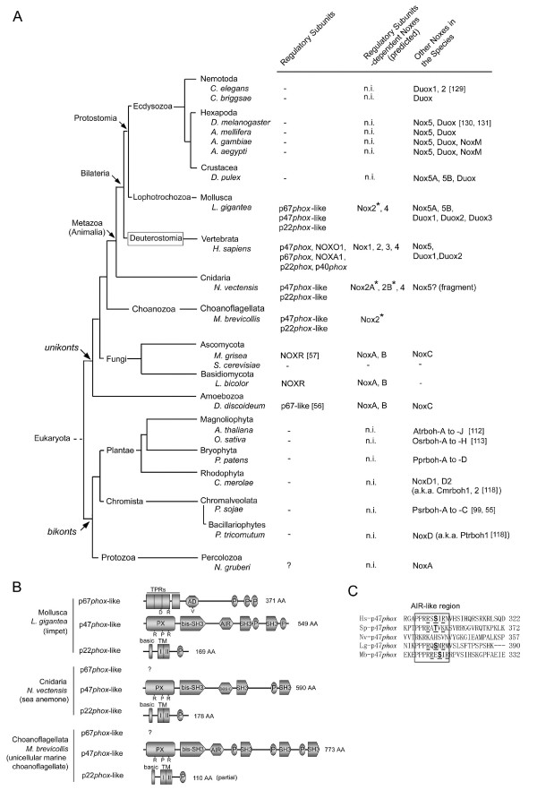
Background: The reactive oxygen-generating NADPH oxidases (Noxes) function in a variety of biological roles, and can be broadly classified into those that are regulated by subunit interactions and those that are regulated by calcium. The prototypical subunit-regulated Nox, Nox2, is the membrane-associated catalytic subunit of the phagocyte NADPH-oxidase. Nox2 forms a heterodimer with the integral membrane protein, p22phox, and this heterodimer binds to the regulatory subunits p47phox, p67phox, p40phox and the small GTPase Rac, triggering superoxide generation. Nox-organizer protein 1 (NOXO1) and Nox-activator 1 (NOXA1), respective homologs of p47phox and p67phox, together with p22phox and Rac, activate Nox1, a non-phagocytic homolog of Nox2. NOXO1 and p22phox also regulate Nox3, whereas Nox4 requires only p22phox. In this study, we have assembled and analyzed amino acid sequences of Nox regulatory subunit orthologs from vertebrates, a urochordate, an echinoderm, a mollusc, a cnidarian, a choanoflagellate, fungi and a slime mold amoeba to investigate the evolutionary history of these subunits.

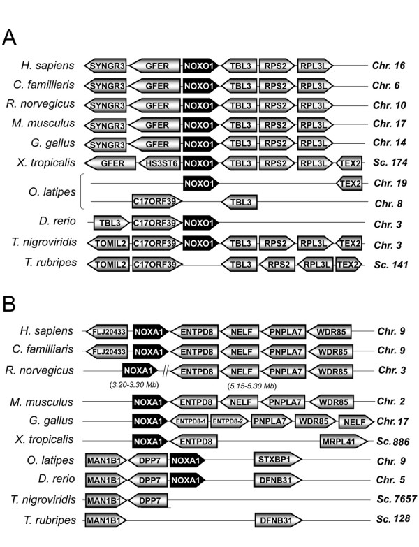
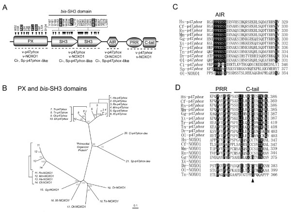
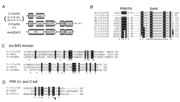
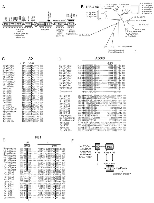
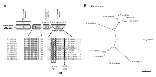
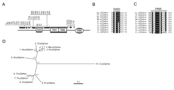
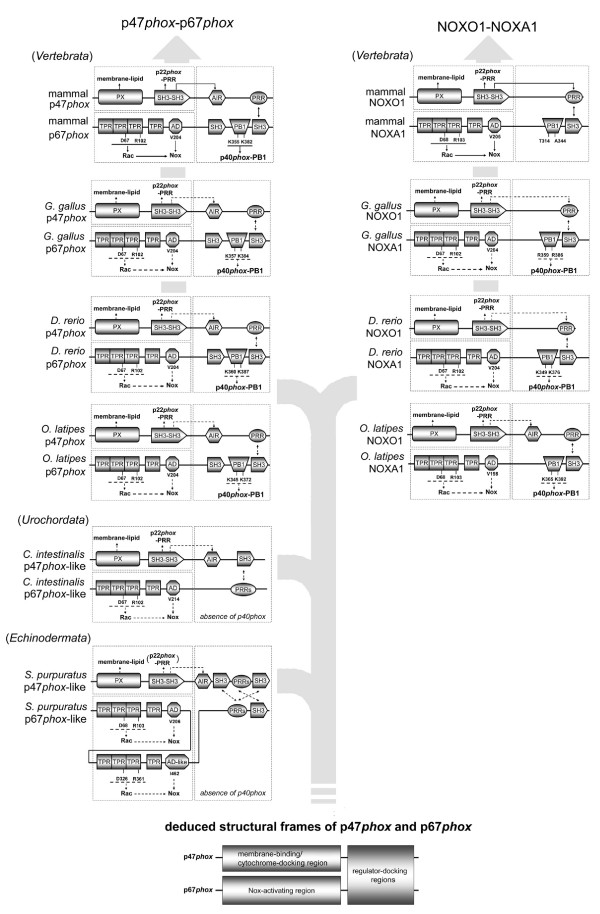
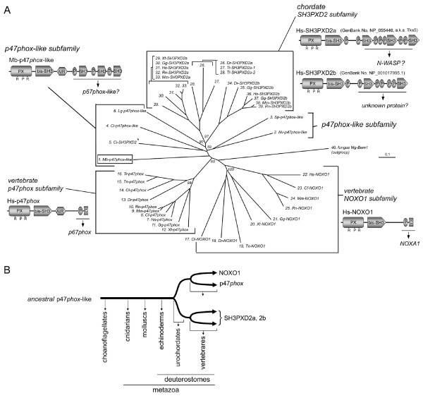
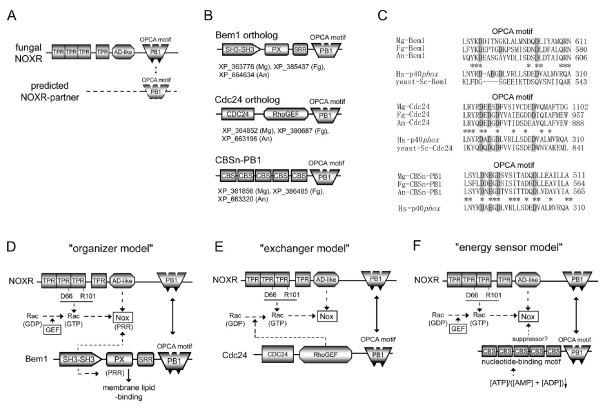
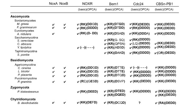
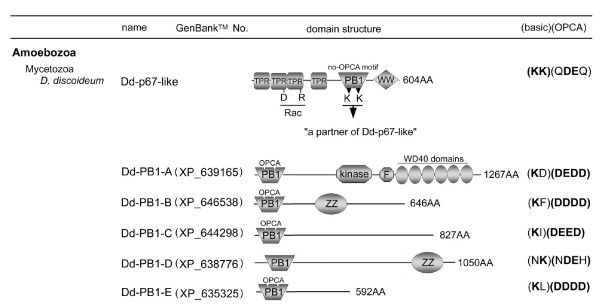
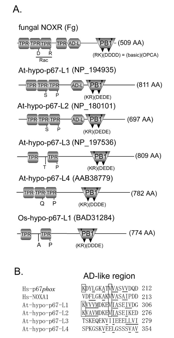
Results: Ancestral p47phox, p67phox, and p22phox genes are broadly seen in the metazoa, except for the ecdysozoans. The choanoflagellate Monosiga brevicollis, the unicellular organism that is the closest relatives of multicellular animals, encodes early prototypes of p22phox, p47phox as well as the earliest known Nox2-like ancestor of the Nox1-3 subfamily. p67phox- and p47phox-like genes are seen in the sea urchin Strongylocentrotus purpuratus and the limpet Lottia gigantea that also possess Nox2-like co-orthologs of vertebrate Nox1-3. Duplication of primordial p47phox and p67phox genes occurred in vertebrates, with the duplicated branches evolving into NOXO1 and NOXA1. Analysis of characteristic domains of regulatory subunits suggests a novel view of the evolution of Nox: in fish, p40phox participated in regulating both Nox1 and Nox2, but after the appearance of mammals, Nox1 (but not Nox2) became independent of p40phox. In the fish Oryzias latipes, a NOXO1 ortholog retains an autoinhibitory region that is characteristic of mammalian p47phox, and this was subsequently lost from NOXO1 in later vertebrates. Detailed amino acid sequence comparisons identified both putative key residues conserved in characteristic domains and previously unidentified conserved regions. Also, candidate organizer/activator proteins in fungi and amoeba are identified and hypothetical activation models are suggested.

Conclusion: This is the first report to provide the comprehensive view of the molecular evolution of regulatory subunits for Nox enzymes. This approach provides clues for understanding the evolution of biochemical and physiological functions for regulatory-subunit-dependent Nox enzymes.


Figures

Figure 1
Figure 1
Relationships between the phylogenic tree of species and the occurrence of Phox-reated regulatory subunits. (A) Summary of the occurrence of Nox-regulatory subunit genes in vertebrates, a urochordate, an echinoderm, fungi, and a slime mold amoeba are shown. Asterisks indicate that the gene is a co-ortholog ancestor of all members of the vertebrate Nox1-3 subgroup. Superscripted letters represent: (a), a partial amino acid sequence predicted from genomic DNA fragment (nucleotides 713622–713565 of GenBank™ No. NW_001471438.1), (b) the gene taxonomy is discussed in the text. (B) Summary of the occurrence of Nox-regulatory subunit genes within Deuterostomia. The number in the fork of each branch indicates the estimated time in millions of years (Mya, millions of years ago) since a given species diverged. A schematic phylogeny and the estimated time of occurrence of species were created from genomic information [62, 125]. Branch lengths are for illustrative purposes and are not proportional to time since divergence.
Figure 2
Figure 2
Syntenies of NOXO1 and NOXA1 genes. Syntenies of the indicated vertebrate NOXO1 (A) and NOXA1 (B) genes are shown. Genes are aligned in columns to illustrate orthology. Chromosome (Chr) or scaffold (Sc) number is indicated on the right. Abbreviations of gene names are shown in Additional file 2.
Figure 3
Figure 3
Identification of highly conserved residues of Nox organizer proteins. Species names were abbreviated as follows: v-, vertebrate; Hs, H. sapiens; Cf, C. familliaris; Rn, R. norvegicus; Mm, M. musculus; Gg, G. gallus; Xt, X. tropicalis, Dr, D. rerio; Tr, T. rubripes; Tn, T. nigroviridis; Ol, O. latipes; Ci, C. intestinalis; Sp, S. purpuratus. (A) Schematic domain structures of Nox organizer are shown; PX, Phox homology; SH3, the Src homology 3; AIR, autoinhibitory region; PRR, proline-rich region; C-tail, C-terminal tail region. Orthologs that possess these domains are shown below the domains. Amino acid residues indicate conserved amino acids in all Nox-organizers. Residue numbers correspond to those of the human p47phox protein sequence. Letters in solid boxes indicate the residues that have been previously proven by mutational analyses to be essential for the human Nox2 activation [24, 68, 126]. (B) Molecular taxonomy was created comparing by the sequence spanning the PX and bis-SH3 domains. (C) Alignment of amino acid sequences of the AIR. Letters in solid boxes indicate residues previously shown to be essential for human Nox2 activation, determined by mutational analyses [20] and also key residues responsible for interactions with the bis-SH3 domain, shown by X-ray crystallography [21]. (D) Alignment of amino acid sequences of the PRR and C-tail regions. Letters in solid boxes indicate the residues that have been shown by mutational analyses to be important for binding to p67phox [18, 72]. An arrow head indicates a residue corresponding to a phosphorylation site of human p47phox [19].
Figure 4
Figure 4
Hypothetical function of T. rubripes C17orf39 protein as a Nox organizer protein. (A) Schematic domain structures of C17orf39 and NOXO1. Names of orthologs that possess domains are shown to the left of structures. T. rubripes C17orf39 was located in scaffold 141 (Sc 141). Abbreviations of species names and most domains are shown in Figure 3; "basic" indicates a lysine/arginine-rich polybasic region. (B) Alignments of the PRR and basic regions of C17orf39. Letters in solid boxes indicate conserved the proline residues in the PRR and lysines and arginines in the basic region. (C) Comparison of bis-SH3 domain among T. rubripes C17orf39, human NOXO1, and human p47phox. (D) Comparison of PRR and C-tail regions among T. rubripes C17orf39, human NOXO1, and human p47phox. Letters in solid boxes indicate the evolutionally conserved residues that have been identified in Figure 3 (C, D). An arrow head indicates a residue corresponding to a phosphorylation site of human p47phox. Identical residues are shown by asterisks (B-D).
Figure 5
Figure 5
Identification of highly conserved residues of Nox activator proteins. Abbreviations of species names are shown in Figure 3. (A) Schematic domains structures of p67phox are shown; TPR, the tetratricopeptide repeats; AD, activation domain; ADSIS, AD-SH3 Intervening Sequence; PB1, Phox/Bem 1; OPCA motif, OPR/PC/AID motif. Abbreviations of other domain names are given in Figure 3. Names of orthologs that possess certain domains are shown below the domains. Amino acid residues indicate conserved amino acids in all indicated proteins, and the residue numbers correspond to that of human p67phox.An asterisk indicates that fungal NOXR possess the OPCA motif. Letters in solid boxes indicate residues previously proven by mutational analyses [27] and by X-ray crystallographic structure analysis [28] to be important for binding to Rac (TRP) or p47phox (SH3-B). (B) Molecular taxonomy was built by the length corresponding to the TPR and AD domains. Species names are abbreviated as shown in Figure 3 and as follows: An, A. nidulans; Mg, M. grisea; Fg, F. graminearum. (C-E) Alignments of the AD, ADSIS, and PB1 domains are shown. (C) Letters in a solid box indicate residues that have been proven by mutational analyses to be essential for activation of human Nox2 [30]. Letters in a gray box indicate basic residues that might correlate with a functional PB1 domain. (D) Amino acid residues identical among vertebrate p67phox ortholog sequences are indicated by asterisks. Letters in gray boxes indicate amino acid residues conserved in the putative ADSIS region. (E) Letters in solid boxes indicate the key basic residues required to bind to another PB1 domain, as shown by X-ray crystallographic structural analysis [32]. K355 and K382 indicate lysine residues of the human p67phox. "β1" and "α1" indicate the first β-sheet and the first α-helical structures of PB1 domain [32]. (F) Proposed model for a cooperation of a basic residue of AD (K196 of the human p67phox sequence) with a functional PB1 domain.
Figure 6
Figure 6
Identification of highly conserved residues of p40phox. (A) A schematic structure of p40phox domains is shown; proline-basic region, P-basic. Abbreviations of domain names are described in legends to other Figures. Letters in gray boxes indicate highly conserved proline and basic amino acid residues in the P-basic region. Letters in solid boxes indicate key acidic residues of OPCA motif that have been demonstrated by X-ray crystallographic structure [32]. K355 and K382 indicate two residues of human p67phox that interact with the OPCA motif. (B) Molecular taxonomy was generated by the alignment of amino acid sequences of PX domain. Species name abbreviations are given in Figure 3.
Figure 7
Figure 7
Identification of highly conserved residues of p22phox. (A) Schematic domains structures of p22phox are shown; TM1 and TM2 represent the predicted 1st and 2nd transmembrane region; PRR, proline-rich region; basic, a lysine- and arginine-rich conserved domain. Indicated above the schematic structure are putative key amino acid residues that are conserved in p22phox orthologs, and the residue numbers correspond to those of human p22phox. Detailed alignment of the polybasic region (B) and the PRR (C) are shown. Letters in solid boxes indicate the residues that have been proven by mutational analyses to be important for binding to p47phox and/or activation of human Nox2 [53, 75, 127, 128]. (D) Molecular taxonomy was generated based on the alignment of amino acid sequences of full-length p22phox. Species name abbreviations are given in Figure 3.
Figure 8
Figure 8
Evolution of domains of p47phox /NOXO1 and p67phox /NOXA1 orthologs. Shown is a schematic representation of identified domains and known or predicted inter- and intra-molecular interactions among p47phox/NOXO1 and p67phox/NOXA1 orthologs in "mammals" (H. sapiens), G. gallus, D. rerio, O. latipes, C. intestinalis, and S. purpuratus. Solid lines indicate interactions that have been demonstrated experimentally. Broken lines indicate predicted or proposed interactions based on sequences analysis and correspondence to experimentally demonstrated interactions in other species. The inferior diagram indicates deduced generalized structural features of p47phox- and p67phox-like proteins. Abbreviations of domain names are given in legends to other Figures.
Figure 9
Figure 9
Relationship between the evolution of eukaryotes and the occurrence of Phox -related regulatory subunits. (A) A phylogeny of eukaryotes was created based largely on genetic information [90, 100]. Branch lengths are for illustrative purposes and are not proportional to phylogenetic distances of groups. Details of deusterostomes are shown in Figure 1. "n.i." indicates none identified or reported. "co-ortho. of v-Nox1-3" indicates that the gene is a co-ortholog ancestor of all members of the vertebrate Nox1-3 subgroup. The number in a parenthesis (e.g. [129, 130, 131]) indicates a reference number. (B) Predicted domains structures of newly identified putative regulatory subunit-like proteins are shown; P, proline-rich region. The sequences are provided in Additional file 7. Abbreviations of domain names are described in legends to other Figures. (C) Sequences of putative AIR-like region are shown. Underlines indicate residues that agree with a consensus sequence of protein kinase C substrate. Bold letters indicate predicted phosphorylation sites in AIR-like regions. Abbreviations: Hs, H. sapiens; Sp, S. purpuratus; Nv, N. vectensis; Lg, L. gigantea; Mb, M. brevicollis.
Figure 10
Figure 10
Evolution of a vast family of p47phox proteins. (A) Molecular taxonomy of the p47phox and related proteins is shown. The sequences of SH3PXD2 orthologs are provided in Additional file 10. Predicted domains structures of H. sapiens (p47phox, NOXO1, SH3PXD2a, 2b) and M. brevicollis (p47phox-like) proteins are shown near to each subfamily: P, proline-rich region; C, C-tail region. An asterisk indicates the appearance of a C. intestinalis SH3PXD2 branch from a common root of the p47phox-like protein. Abbreviations of species names and other domains are given in other Figures. (B) A schematic drawing to show a hypothetical evolution of an expanded p47phox-like gene family. Branches are for illustrative purposes and are not proportional to rates of divergence.
Figure 11
Figure 11
Predicted binding partners of fungal NOXR and activation models of fungal Nox. (A) A hypothetical domain structure of a predicted NOXR partner is shown. (B) Domain structures of fungal proteins with PB1 domains meeting criteria for possible NOXR-partners are shown, along with their GenBank™ accession numbers. Abbreviations: An, A. nidulans; Mg, M. grisea; Fg, F. graminearum. (C) Aligned OPCA motifs of candidate NOXR partners are compared with the corresponding region in p40phox and the yeast isolog (the first and second alignments). Amino acid residues conserved among fungal ortholog sequences are indicated by asterisks. (D) A proposed model for activation of fungal Nox by NOXR and Bem1 protein. (E) A proposed model for activation of fungal Nox by NOXR and Cdc24. (F) A proposed model for activation of fungal Nox by CBSn-PB1 proteins. RhoGEF, Rho guanine nucleotide exchange factor; CBS, cystathionine beta-synthase; Abbreviations of other domain names are described in legends to other Figures.
Figure 12
Figure 12
Conservations of NOXR, Bem1, Cdc24, and CBSn-PB1 genes and characteristic residues of PB1 domains in fungal genomes. (A) "basic" and "OPCA" in parenthesis indicate key residues that are required to form a dimer between two PB1 domains; two "basic" residues and four in "OPCA" motif residues correspond to Lys-355 and Lys-382 of human p67phox and Asp-289, Glu-291, Asp-293 and Asp-302 of human p40phox, respectively. Divisions of Fungi are shown in bold.
Figure 13
Figure 13
Predicted candidate binding partners of D. discoideum p67-like protein. Predicted domain structures of Dd-p67-like protein and candidates of binding partners are shown. "basic" and "OPCA" in parenthesis indicate six characteristic residues of PB1 domain as shown in Figure 12. Abbreviations: kinase, protein serine/threonine kinase domain; F, F-box; WD40, WD40 repeat; WW, WW domain; ZZ, ZZ type zinc-finger.
Figure 14
Figure 14
Predicted p67phox- like proteins in higher plants. (A) Predicted domain structures of hypothetical p67phox-like proteins (At-hypo-p67-L1 to -L4, Os-hypo-p67-L1) of A. thaliana and O. sativa, and a fungal F. graminearum (Fg-) NOXR are shown, along with their GenBank™ accession numbers. "basic" and "OPCA" in parenthesis indicate six characteristic residues of PB1 domains as shown in Figure 12. "D" and "R" below the TPR region of Fg-NOXR represent two conserved amino acid residues that are important for binding to Rac as indicated in Figure 5. (B) Alignments of the putative AD-like regions are shown (see details in Additional file 17). Boxes indicate lysine and valine residues corresponding to the Lys-196 and Val-204 of human p67phox, respectively. Underlines in alignment indicates amino acid with a hydropholic side group.

Similar articles

Cited by

References

    1. Kawahara T, Quinn MT, Lambeth JD. Molecular evolution of the reactive oxygen-generating NADPH oxidase (Nox/Duox) family of enzymes. BMC Evolutionary Biology. 2007;7 - PMC - PubMed
    1. Bedard K, Lardy B, Krause KH. NOX family NADPH oxidases: Not just in mammals. Biochimie. 2007;89:1107–1112. doi: 10.1016/j.biochi.2007.01.012. - DOI - PubMed
    1. Vignais PV. The superoxide-generating NADPH oxidase; structural aspects and activation mechanism. Cellular and Molecular Life Sciences. 2002;59:1428–1459. doi: 10.1007/s00018-002-8520-9. - DOI - PMC - PubMed
    1. Heyworth PG, Cross AR, Curnutte JT. Chronic granulomatous disease. Current Opinion in Immunology. 2003;15:578–584. doi: 10.1016/S0952-7915(03)00109-2. - DOI - PubMed
    1. Lambeth JD. Nox enzymes, ROS, and chronic disease: an example of antagonistic pleiotropy. Free Radical Biology and Medicine. 2007;43:332–347. doi: 10.1016/j.freeradbiomed.2007.03.027. - DOI - PMC - PubMed

Publication types

Substances