. 2007 Nov 9;373(5):1184-97.
doi: 10.1016/j.jmb.2007.08.050. Epub 2007 Aug 28.

Kinetic analysis of the slow skeletal myosin MHC-1 isoform from bovine masseter muscle

Affiliations

Kinetic analysis of the slow skeletal myosin MHC-1 isoform from bovine masseter muscle

M J Bloemink et al. J Mol Biol. .

Abstract

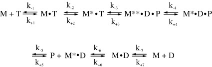
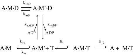
Several heavy chain isoforms of class II myosins are found in muscle fibres and show a large variety of different mechanical activities. Fast myosins (myosin heavy chain (MHC)-II-2) contract at higher velocities than slow myosins (MHC-II-1, also known as beta-myosin) and it has been well established that ADP binding to actomyosin is much tighter for MHC-II-1 than for MHC-II-2. Recently, we reported several other differences between MHC-II isoforms 1 and 2 of the rabbit. Isoform II-1 unlike II-2 gave biphasic dissociation of actomyosin by ATP, the ATP-cleavage step was significantly slower for MHC-II-1 and the slow isoforms showed the presence of multiple actomyosin-ADP complexes. These results are in contrast to published data on MHC-II-1 from bovine left ventricle muscle, which was more similar to the fast skeletal isoform. Bovine MHC-II-1 is the predominant isoform expressed in both the ventricular myocardium and slow skeletal muscle fibres such as the masseter and is an important source of reference work for cardiac muscle physiology. This work examines and extends the kinetics of bovine MHC-II-1. We confirm the primary findings from the work on rabbit soleus MHC-II-1. Of significance is that we show that the affinity of ADP for bovine masseter myosin in the absence of actin (represented by the dissociation constant K(D)) is weaker than originally described for bovine cardiac myosin and thus the thermodynamic coupling between ADP and actin binding to myosin is much smaller (K(AD)/K(D) approximately 5 instead of K(AD)/K(D) approximately 50). This may indicate a distinct type of mechanochemical coupling for this group of myosin motors. We also find that the ATP-hydrolysis rate is much slower for bovine MHC-II-1 (19 s(-1)) than reported previously (138 s(-1)). We discuss how this work fits into a broader characterisation of myosin motors from across the myosin family.


Figures

Figure 1
Figure 1
SDS–polyacrylamide electrophoresis gel of BMS1 purification. Samples were run on a SDS–15% gel with glycine/Tris/SDS running buffer. Lane 1, bovine masseter myosin (BMMyosin) with two light chains. Lane 2, BMS1 after chymotrypsin digestion and column purification. Lane 3, molecular mass marker.
Figure 2
Figure 2
Rate of association of ATP or ADP with BMS1. (a) The protein fluorescence changes observed on mixing 0.5 μM BMS1 with 25 μM ATP or 400 μM ATP at 20 °C. For the reaction with 25 μM ATP the best-fit double exponential is superimposed with kobs = 19.6 s−1F +11.8%) and 2.5 s−1F +4%) for the fast and medium/slow phase, respectively. The fluorescence of the reaction with 400 μM ATP could best be fitted to a triple exponential with kobs = 111 s−1F +10%), kobs = 13.5 s−1 ((ΔF +3.7%) and kobs = 2 s−1F +1%) for the fast, medium and slow phase, respectively. (b) Dependence of kobs on ATP concentration. At high ATP-concentrations, kobs saturates at 117 s−1 for the fast phase (▪), at 18 s−1 for the medium phase (•) and 2.0 s−1 for the slow phase (▴). At low ATP concentrations the data fit a straight line with slope (K1k+2) = 1.5 × 106 M−1s−1 for the fast phase. (c) Dependence of kobs on ADP concentration, with a saturation value for kobs = 26 s−1. The maximal rate of ADP-binding represents k+6 +k-6 (kmax,ADP) = 26 s−1 and K7 = K0.5,ADP = 21 μM. The intercept yields k+6 (=k-D) = 1.4(±0.5) s−1. The second-order constant of ADP binding k-6/K7 (=k+D) is defined by kmax,ADP/K0.5,ADP = 1.24 × 106 M−1s−1, resulting in KD (k-D/k+D) = 1.2 μM.
Figure 3
Figure 3
Kinetics of MgATP hydrolysis by BMS1 using rapid quench-flow at 20 °C (100 mM KCl, 20 mM Mops, 5 mM MgCl2(pH 7)). 5 μM BMS1 is first mixed with 50 μM MgATP and after variable time quenched with 6.25% TCA The ADP/total nucleotide ratio is plotted as a function of time and can be fitted to a single exponential, resulting in a time constant t for the initial burst (t = 52 ms), resulting in a kobs of 19 s−1.
Figure 4
Figure 4
Displacement of ADP from BMS1-ADP by addition of excess ATP. (a) and (b) Protein fluorescence changes observed on mixing 200 μM ATP with 1.0 μM BMS1 and 0.4 μM ADP. Comparison of the sum of two exponentials (a) or three exponentials (b) shows that the data fit best to a sum of three exponentials. The measured values of the kobs in (b) were 68 s−1 (fast phase, ΔF +8.9%), 9.6 s−1 (medium phase, ΔF +2.2%) and 0.9 s−1 (slow phase, ΔF +6%), respectively. (c) Dependence of the relative amplitudes of the three exponentials on ADP concentration. The data are fitted to hyperbolae with a Kd of 2.6 μM (fast phase, ▪), 3.0 μM (medium phase, •) and 3.0 μM (slow phase, ▴).
Figure 5
Figure 5
ATP-induced dissociation of pyr-acto–BMS1. (a) The pyrene fluorescence changes observed on mixing 0.5 μM BMS1 with 200 μM ATP at 20 °C. The best-fit double exponential is superimposed with kobs = 278 s−1F +77%) and 58 s−1F +10%) for the fast and slow phase, respectively. (b) Dependence of kobs on [ATP]. At high ATP-concentrations, kobs saturates at ∼1100 s−1 for the fast phase and remains constant at 56(±13) s−1 for the slow phase. (c) At low [ATP] the data of the fast phase fit a straight line with slope (K1k+2) = 1.23 × 106 M−1s−1.
Figure 6
Figure 6
ATP-induced dissociation of pyr-acto–BMS1 in the presence of ADP. 0.25 μM phalloidin-stabilized pyrene-labelled actin was incubated with an equimolar amount of BMS1 before mixing with variable concentrations of ADP and 50 μM ATP (a) or 200 μM ATP (b). The data could be fitted against a single exponential (a) or a double exponential (b) and the resulting kobs were fitted toa hyperbola, resulting in an apparent affinity (KAD) forADP (a) KAD = 9.6(±1.4) μM (20 °C) and (b) KAD = 6.8(±1.7) μM (▪, fast phase) and KAD = 6.6(±1.9) μM (•, slow phase).
Figure 7
Figure 7
ATP-induced dissociation of BMS1-actin in the presence of ADP. (a) Pyrene fluorescence changes observed on mixing 16 mM MgATP with 1 μM BMS1-actin in the presence of 150 μM ADP at 20 °C. The best-fit double exponential is superimposed with kobs = 94 s−1F+55%) and 9.6 s−1F +10%) for the fast and slow phase, respectively. (b) Temperature dependence of ATP-induced dissociation of BMS1-actin in the presence of ADP with kobs (fast (▪) and slow (•) phase) as a function of temperature. From the slope the activation energy can be calculated. Ea = 106 kJmol−1 and 98 kJmol−1 for the fast and the slow phase, respectively.
Figure 8
Figure 8
Titration of actin with BMS1. 30 nM phalloidin-stabilised pyrene-labelled actin was incubated with various amounts of BMS1 before mixing with 500 μM ATP in the presence of 500 μM ADP (▪) or with 20 μM ATP without ADP (□). In both cases the reaction was well described by a single exponential and the amplitude increased with increasing [BMS1]. The best fit to the quadratic equation describing the binding isotherm (see Materials and Methods) gave a Kd of 7 nM and 37 nM in the absence and presence of ADP, respectively (concentrations used here are before mixing).
Scheme 1
Scheme 1
Proposed reaction scheme for the interaction of BMS1 with nucleotides (T, ATP; D, ADP) as described.
Scheme 2
Scheme 2
Proposed reaction scheme for the interaction of BMS1 with nucleotides (T, ATP; D, ADP) in the presence of actin as proposed for myosin I (A represents actin and M is myosin S1).

Similar articles

Cited by

References

    1. De La Cruz E.M., Ostap E.M. Relating biochemistry and function in the myosin superfamily. Curr. Opin. Cell Biol. 2004;16:61–67. - PubMed
    1. Geeves M.A., Holmes K.C. The molecular mechanism of muscle contraction. Adv. Protein Chem. 2005;71:161–193. - PubMed
    1. Reggiani C., Bottinelli R., Stienen G.J. Sarcomeric myosin isoforms: fine tuning of a molecular motor. News Physiol. Sci. 2000;15:26–33. - PubMed
    1. Timson D.J. Fine tuning the myosin motor: the role of the essential light chain in striated muscle myosin. Biochimie. 2003;85:639–645. - PubMed
    1. Schiaffino S., Reggiani C. Molecular diversity of myofibrillar proteins: gene regulation and functional significance. Physiol. Rev. 1996;76:371–423. - PubMed

Publication types