Review
. 2007 Nov 10;59(13):1329-39.
doi: 10.1016/j.addr.2007.08.007. Epub 2007 Aug 14.

Cell responses to the mechanochemical microenvironment--implications for regenerative medicine and drug delivery

Affiliations
Review

Cell responses to the mechanochemical microenvironment--implications for regenerative medicine and drug delivery

Florian Rehfeldt et al. Adv Drug Deliv Rev. .

Abstract

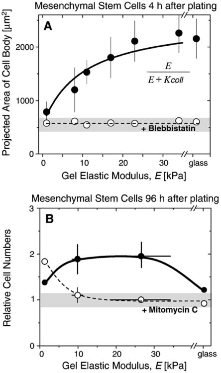
Soft-tissue cells are surprisingly sensitive to the elasticity of their microenvironment, suggesting that traditional culture plastic and glass are less relevant to tissue regeneration and chemotherapeutics than might be achieved. Cells grown on gels that mimic the elasticity of tissue reveal a significant influence of matrix elasticity on adhesion, cytoskeletal organization, and even the differentiation of human adult derived stem cells. Cellular forces and feedback are keys to how cells feel their mechanical microenvironment, but detailed molecular mechanisms are still being elucidated. This review summarizes our initial findings for multipotent stem cells and also the elasticity-coupled effects of drugs on cancer cells and smooth muscle cells. The drugs include the contractility inhibitor blebbistatin, the proliferation inhibitor mitomycin C, an apoptotis-inducing antibody against CD47, and the translation inhibitor cycloheximide. The differential effects not only lend insight into mechano-sensing of the substrate by cells, but also have important implications for regeneration and molecular therapies.


Figures

Fig. 1
Fig. 1
Cells on elastic substrates that model tissue elasticity. A) Sketch of a model in vitro environment of a cell on a substrate of elasticity E, coated with ligands that are specifically recognized by cell adhesion receptors. Force sensing and transduction is mediated by these contacts. Biochemical stimuli are also provided by factors in the surrounding media. B) Elasticity of various solid tissues, and blood as a “fluid tissue”.
Fig. 2
Fig. 2
Cells exert force on the underlying substrate. (Top) A 3T3 fibroblast on a silicone rubber substrate deforms the film, causing wrinkles. Scale bar is 10 μm. (Bottom) Group of chick heart fibroblasts forming a more complex wrinkle pattern showing that strain transmission through the substrate propagates to neighboring cells. Scale bar is 100 μm. (Reprinted by permission from Macmillan Publishers Ltd: Nature (Harris, A.K., D. Stopak, and P. Wild, Fibroblast Traction as a Mechanism for Collagen Morphogenesis. Nature, 1981 290(5803): p. 249–251.), copyright (1981)).
Fig. 3
Fig. 3
Measurement of micro-elasticity of matrices by AFM. A) A cantilever with a pyramidal tip (opening angle α) is translated down (Δz) into the sample causing a deflection d monitored by the photodiode, yielding the indentation δzd. The required force F=k Δd is defined by the spring constant k multiplied by the deflection d. B) The Young's modulus E is determined by analyzing the resulting force-indentation curves with a modified Hertz model. Black curve denotes data points; red solid line is the best fit resulting from the modified Hertz model. (For interpretation of the references to colour in this figure legend, the reader is referred to the web version of this article.)
Fig. 4
Fig. 4
Mesenchymal stem cell (MSC) differentiation on elastic substrates. A) Morphology of naive, low passage MSCs (upper panel) 24 h after plating on PA gels of different stiffness closely matches that of cell lineages found within each microenvironment. Naive MSCs are initially small and round but a dominant fraction indicated here (lower panel) develops increasingly branched, spindle, or polygonal shapes within days of plating when grown on matrices with respective elasticities of 1 kPa, 11 kPa, and 34 kPa. Results for mitomycin C treated cells are shown with diagonally-hatched bars. Scale bar is 20 μm. B) Differentiation of MSCs directed by substrate elasticity elucidated by key marker proteins. The neuronal cytoskeletal marker (β3 tubulin is expressed in branches (arrows) of initially naive MSCs (>75%) and only on soft, neurogenic matrices (first row). The muscle transcription factor MyoD is up-regulated and nuclear localized (arrow) only in MSCs on myogenic matrices (second row). The osteoblast transcription factor CBFα1 (arrow) is likewise expressed only on stiff osteogenic substrates (third row). Scale bar is 5 μm.
Fig. 5
Fig. 5
Interplay of adhesion, force-sensing, and signal transduction. Integrins bind extracellular matrix ligands and link to the cytoskeleton. Signaling proteins such as Rac and Rho are up and down regulated coupled to myosin II-based contractility, thereby influencing local and global adhesion.
Fig. 6
Fig. 6
MSC spreading, proliferation, and drug responses. A) Area of MSC cell body as measured on PA gels of different stiffnesses and on glass. Cell area increases with stiffness unless treated with the myosin II inhibitor, blebbistatin. The solid line shows the best fit using the function shown as inset yielding K coll=10 kPa that agrees well with values determined for smooth muscle cells. B) MSC cell numbers relative to the number plated on elastic PA hydrogels. On soft gels, mitomycin C treated cells (open circles) seem to be more proliferative than the untreated control cells (full grey circles), whereas the behavior reverses on very rigid surfaces and on glass, proliferation is suppressed due to strong adhesion to stiffer substrates.
Fig. 7
Fig. 7
Substrate elasticity influences spreading and antibody-directed drug treatment of cancer cells. (A) Human lung cancer epithelial cells (A549) spread on collagen I coated rigid substrates, but 4 kPa gels induce little cell spreading. Images show viable cells are red-stained with mito-tracker dye, but the dye disperses to cytoplasm and changes to green with cell death. (B) Addition of an apoptosis inducing antibody against CD47 (B6H12) has little effect on the viability of cells on soft gels, but is effective against cells spread on rigid substrates. (For interpretation of the references to colour in this figure legend, the reader is referred to the web version of this article.)
Fig. 8
Fig. 8
Cell area of smooth muscle cells spread on elastic surfaces and glass, with and without ribosome inhibition with cycloheximide (CHX). Treated cells show significant (p<0.01) differences versus control cells only, when grown on stiffer surfaces (34 kPa and rigid glass). Top images show actin (red) and ribosomes (green) that are found throughout the cytoplasm and at the cell edge. Right images show the actin cytoskeleton is usually organized on rigid matrices unless CHX-treated. (For interpretation of the references to colour in this figure legend, the reader is referred to the web version of this article.)

References

    1. Discher DE, Janmey P, Wang YL. Tissue cells feel and respond to the stiffness of their substrate. Science. 2005;310(5751):1139–1143. - PubMed
    1. Hynes RO. Cell adhesion: old and new questions. Trends in Biochemical Sciences. 1999;24(12):M33–M37. - PubMed
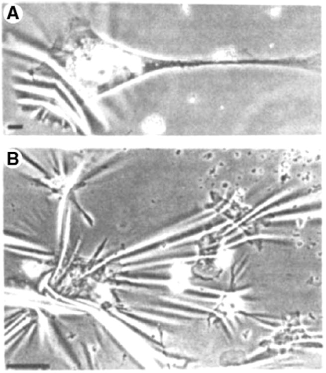
    1. Harris AK, Wild P, Stopak D. Silicone–rubber substrata—new wrinkle in the study of cell locomotion. Science. 1980;208(4440):177–179. - PubMed
    1. Pelham RJ, Wang YL. Cell locomotion and focal adhesions are regulated by substrate flexibility. Proceedings of the National Academy of Sciences of the United States of America. 1997;94(25):13661–13665. - PMC - PubMed
    1. Dembo M, Wang YL. Stresses at the cell-to-substrate interface during locomotion of fibroblasts. Biophysical Journal. 1999;76(4):2307–2316. - PMC - PubMed

Publication types