

Climbing-fibre activation of NMDA receptors in Purkinje cells of adult mice
- PMID: 17901118
- PMCID: PMC2327252
- DOI: 10.1113/jphysiol.2007.141531
Climbing-fibre activation of NMDA receptors in Purkinje cells of adult mice
Abstract
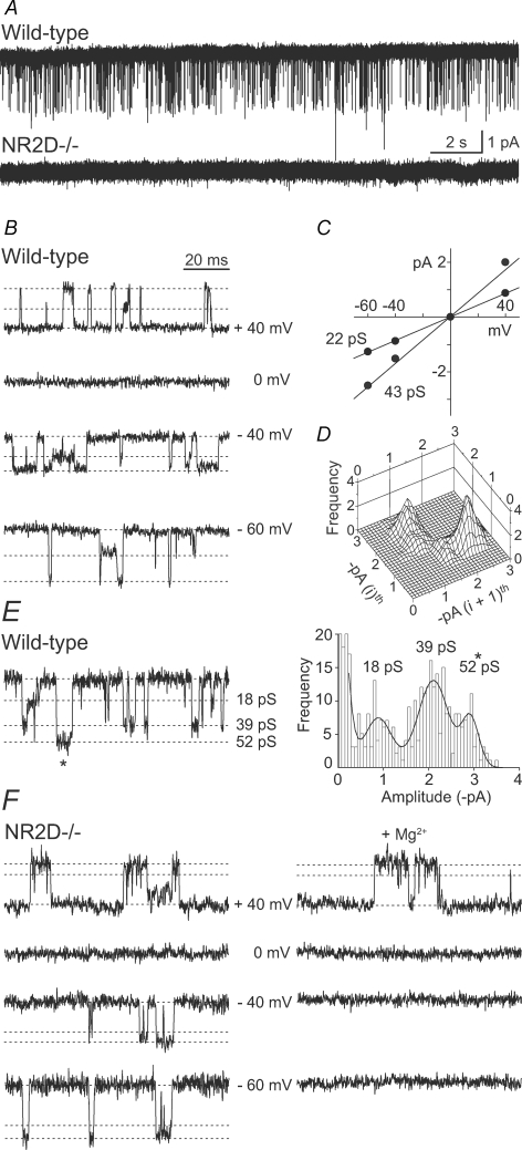
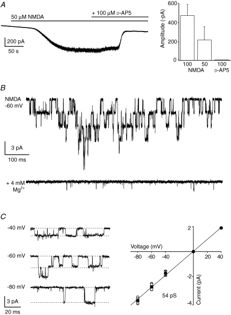
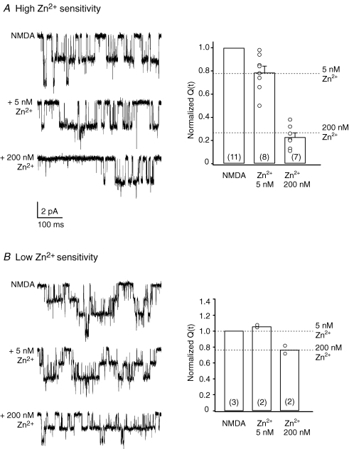
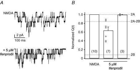
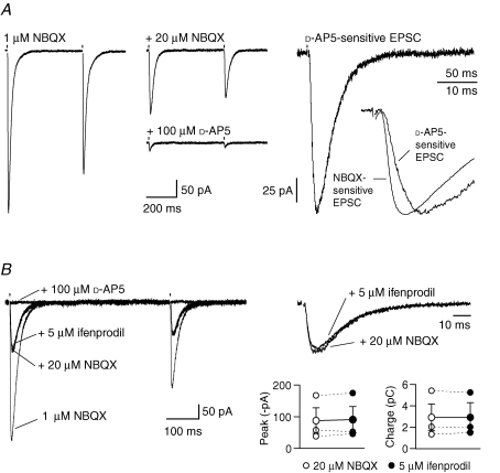
Among principal neurons, adult Purkinje cells have long been considered unusual in lacking functional NMDA receptors. This view has emerged largely from studies on rats, where NMDA receptors are expressed in Purkinje cells of newborn animals, but are lost after 2 weeks. By contrast, immunolabelling data have shown that Purkinje cells from adult mice express multiple NMDA receptor subunits, suggesting a possible species difference. To investigate the presence of functional NMDA receptors in Purkinje cells of mice, and to explore the contribution of different receptor subunits, we made whole-cell and single-channel patch-clamp recordings from Purkinje cells of wild-type and NR2D-/- mice of different ages. Here we report that multiple NMDA receptor subtypes are indeed expressed in Purkinje cells of young and adult mice; in the adult, both NR2A- and NR2B-containing subtypes are present. Furthermore, we show that NMDA receptor-mediated EPSCs can be evoked by climbing fibre stimulation, and appear to be mediated mainly by NR2A-containing receptors.
Figures
Similar articles
-
NMDA receptor contribution to the climbing fiber response in the adult mouse Purkinje cell.J Neurosci. 2007 Oct 3;27(40):10797-809. doi: 10.1523/JNEUROSCI.2422-07.2007. J Neurosci. 2007. PMID: 17913913 Free PMC article.
-
Purkinje cell NMDA receptors assume a key role in synaptic gain control in the mature cerebellum.J Neurosci. 2010 Nov 10;30(45):15330-5. doi: 10.1523/JNEUROSCI.4344-10.2010. J Neurosci. 2010. PMID: 21068337 Free PMC article.
-
Critical period for activity-dependent synapse elimination in developing cerebellum.J Neurosci. 2000 Jul 1;20(13):4954-61. doi: 10.1523/JNEUROSCI.20-13-04954.2000. J Neurosci. 2000. PMID: 10864953 Free PMC article.
-
Characterization of L-homocysteate-induced currents in Purkinje cells from wild-type and NMDA receptor knockout mice.J Neurophysiol. 1999 Nov;82(5):2820-6. doi: 10.1152/jn.1999.82.5.2820. J Neurophysiol. 1999. PMID: 10561449
-
Excitatory synaptic currents in Purkinje cells.Proc Biol Sci. 1990 Aug 22;241(1301):116-21. doi: 10.1098/rspb.1990.0074. Proc Biol Sci. 1990. PMID: 1978337
Cited by
-
Calcium threshold shift enables frequency-independent control of plasticity by an instructive signal.Proc Natl Acad Sci U S A. 2016 Nov 15;113(46):13221-13226. doi: 10.1073/pnas.1613897113. Epub 2016 Oct 31. Proc Natl Acad Sci U S A. 2016. PMID: 27799554 Free PMC article.
-
Presynaptic NR2A-containing NMDA receptors implement a high-pass filter synaptic plasticity rule.Proc Natl Acad Sci U S A. 2009 Aug 18;106(33):14126-31. doi: 10.1073/pnas.0904284106. Epub 2009 Aug 4. Proc Natl Acad Sci U S A. 2009. PMID: 19666514 Free PMC article.
-
Contribution of plasma membrane Ca ATPase to cerebellar synapse function.World J Biol Chem. 2010 May 26;1(5):95-102. doi: 10.4331/wjbc.v1.i5.95. World J Biol Chem. 2010. PMID: 21540995 Free PMC article.
-
Region-specific role for GluN2B-containing NMDA receptors in injury to Purkinje cells and CA1 neurons following global cerebral ischemia.Neuroscience. 2015 Jan 22;284:555-565. doi: 10.1016/j.neuroscience.2014.10.033. Epub 2014 Oct 24. Neuroscience. 2015. PMID: 25450957 Free PMC article.
-
GluN1 splice variant control of GluN1/GluN2D NMDA receptors.J Physiol. 2012 Aug 15;590(16):3857-75. doi: 10.1113/jphysiol.2012.234062. Epub 2012 May 28. J Physiol. 2012. PMID: 22641781 Free PMC article.
References
-
- Auger C, Attwell D. Fast removal of synaptic glutamate by postsynaptic transporters. Neuron. 2000;28:547–558. - PubMed
-
- Cull-Candy S, Brickley S, Farrant M. NMDA receptor subunits: diversity, development and disease. Curr Opin Neurobiol. 2001;11:327–335. - PubMed
-
- Dingledine R, Borges K, Bowie D, Traynelis SF. The glutamate receptor ion channels. Pharmacol Rev. 1999;51:7–61. - PubMed
-
- Dzubay JA, Otis TS. Climbing fiber activation of metabotropic glutamate receptors on cerebellar Purkinje neurons. Neuron. 2002;36:1159–1167. - PubMed
Publication types
MeSH terms
Substances
Grants and funding
LinkOut - more resources
Full Text Sources
Molecular Biology Databases