. 2008 Jan;22(1):167-75.
doi: 10.1210/me.2007-0140. Epub 2007 Sep 27.

Fibroblast growth factor receptor 1 (FGFR1) tyrosine phosphorylation regulates binding of FGFR substrate 2alpha (FRS2alpha) but not FRS2 to the receptor

Affiliations

Fibroblast growth factor receptor 1 (FGFR1) tyrosine phosphorylation regulates binding of FGFR substrate 2alpha (FRS2alpha) but not FRS2 to the receptor

Yongyou Zhang et al. Mol Endocrinol. 2008 Jan.

Abstract

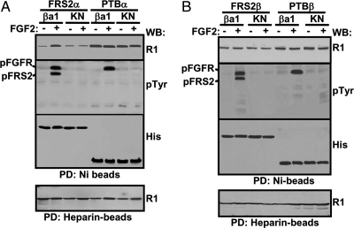
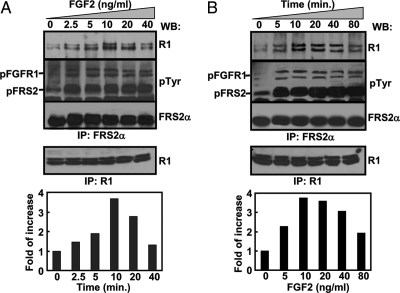
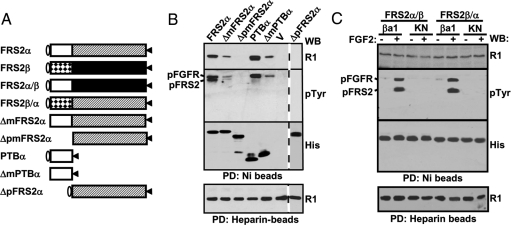
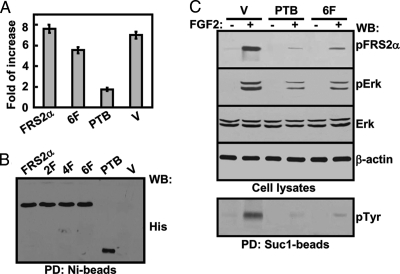
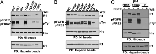
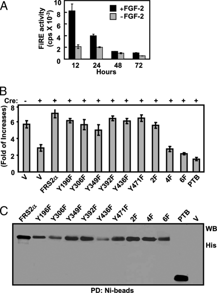
Binding of the fibroblast growth factor (FGF) to the FGF receptor (FGFR) tyrosine kinase leads to receptor tyrosine autophosphorylation as well as phosphorylation of multiple downstream signaling molecules that are recruited to the receptor either by direct binding or through adaptor proteins. The FGFR substrate 2 (FRS2) family consists of two members, FRS2alpha and FRS2beta, and has been shown to recruit multiple signaling molecules, including Grb2 and Shp2, to FGFR1. To better understand how FRS2 interacted with FGFR1, in vivo binding assays with coexpressed FGFR1 and FRS2 recombinant proteins in mammalian cells were carried out. The results showed that the interaction of full-length FRS2alpha, but not FRS2beta, with FGFR1 was enhanced by activation of the receptor kinase. The truncated FRS2alpha mutant that was comprised only of the phosphotyrosine-binding domain (PTB) bound FGFR1 constitutively, suggesting that the C-terminal sequence downstream the PTB domain inhibited the PTB-FGFR1 binding. Inactivation of the FGFR1 kinase and substitutions of tyrosine phosphorylation sites of FGFR1, but not FRS2alpha, reduced binding of FGFR1 with FRS2alpha. The results suggest that although the tyrosine autophosphorylation sites of FGFR1 did not constitute the binding sites for FRS2alpha, phosphorylation of these residues was essential for optimal interaction with FRS2alpha. In addition, it was demonstrated that the Grb2-binding sites of FRS2alpha are essential for mediating signals of FGFR1 to activate the FiRE enhancer of the mouse syndecan 1 gene. The results, for the first time, demonstrate the specific signals mediated by the Grb2-binding sites and further our understanding of FGF signal transmission at the adaptor level.


Figures

Figure 1
Figure 1
Tyrosine Phosphorylation Regulates the Interaction of FGFR1 with Full-Length FRS2α But Not FRS2β and PTB Domains 293T cells transiently coexpressing FRS2α (A) or FRS2β (B) with the indicated FGFR1 constructs were lysed with or without prior treatment of FGF2 for 10 min. The lysates were rocked with Ni- or heparin-agarose beads, and the specifically bound fractions were Western blotted with the indicated antibody. ba1, FGFR1βa1 isoform; KN, kinase-inactive mutant of FGFR1βa1; PTBα, PTB domain of FRS2α; PTBβ, PTB domain of FRS2β; pFGFR, phosphorylated FGFR1; pFRS2, phosphorylated FRS2; R1, anti-FGFR1 antibody; His, anti-His-tag antibody; pTyr, anti-phosphotyrosine antibody; PD, pull-down; WB, Western blot.
Figure 2
Figure 2
The Endogenous FGFR1/FRS2α Interaction Induced by FGF2 Is Dosage and Time Dependent The serum-starved TRAMP C2 cells were treated with FGF2 at the indicated concentrations for 10 min at 37 C (A) or treated with 10 ng/ml FGF2 for the indicated times (B) before being lysed. The FGFR1-FRS2α complexes pulled down from the cell lysates were Western blotted with the indicated antibodies. The bands representing FGFR1 were quantitated with a densitometer. The data were expressed as fold of increases from untreated samples. pFGFR, Phosphorylated FGFR1; pFRS2, phosphorylated FRS2; R1, anti-FGFR1 antibody; His, anti-His-tag antibody; pTyr, anti-phosphotyrosine antibody; Shp2, anti-Shp2 antibody; PD, pull-down; WB, Western blot.
Figure 3
Figure 3
The FRS2α C-Terminal Domain Is Required for Phosphorylation-Regulated FGFR1/FRS2α Interaction A, Schematic of FRS2 mutants. Oval, The conserved myristylation sequence; open box, PTBα; dotted box, PTBβ; hatched box, the C-terminal sequence of FRS2α; solid box, the C-terminal sequence of FRS2β; triangles, the hexahistidine tag. B, The Sf9 expressed FRS2α mutants and FGFR1 kinase were incubated with Ni- or heparin-agarose beads as described, and the specifically bound fractions were subject to Western analyses as described. C, The 293 cells cotransfected with the assigned FGFR1 and FRS2 mutants were treated with 10 ng/ml FGF2 for 10 min where indicated before being lysed with the lysis buffer. The cell lysates were incubated with Ni- and heparin-beads as specified, and the specifically bound fractions were subjected to Western analyses as described. βa1, FGFR1βa1 isoform; KN, kinase-inactive mutant of FGFR1βa1; pFGFR, phosphorylated FGFR1; pFRS2, phosphorylated FRS2; R1, anti-FGFR1 antibody; His, anti-His-tag antibody; pTyr, anti-phosphotyrosine antibody; PD, pull-down; WB, Western blot; dotted lines indicate that the data were put together from different gels.
Figure 4
Figure 4
The Dominant-Negative Inhibition Activity of the PTB Domain A, NIH 3T3 cells cotransfected with the indicated FRS2α mutants and the FiRE-luciferase reporter were stimulated with FGF2 overnight where indicated. The cells were then lysed, and the luciferase activity was measured. After being normalized with the cell number, the data derived from three experiments are presented as fold of increases in response to FGF2 treatments. B, The expressed FRS2α mutants in the same cells as in A were enriched with Ni-beads and Western analyzed with the anti-His-tag antibody. C, NIH 3T3 cells transfected with the indicated FRS2α mutants were treated with FGF2 for 10 min where indicated before being lysed with the RIPA buffer. The cell lysates were Western analyzed with the indicated antibodies. Bottom panel, the same cell lysates were incubated with Suc13-beads, and the specifically bound fractions were Western analyzed with the anti-phosphotyrosine antibody. His, Anti-His-tag antibody; Erk, anti-Erk antibody; pErk, anti-phosphorylated Erk; pFRS2α, anti-phospho-FRS2α antibody; pTyr, anti-phosphotyrosine antibody; PTB, PTB domain of FRS2α; 2F, FRS2α2F mutant; 4F, FRS2α 4 F mutant; 6F, FRS2α mutant with substitutions of all six phosphorylation sites with phenylalanine residues; V, vector only; PD, pull-down; WB, Western blot.
Figure 5
Figure 5
Tyrosine Phosphorylations of FGFR1 But Not FRS2α Promote Interaction of FGFR1 and FRS2α A and B, Sf9 cells coexpressing FRS2α and FGFR1 mutants (A) or FGFR1 and FRS2α mutants (B) were pulled down with Ni- or heparin-beads as specified for Western analysis with the indicated antibodies. C, 293T cells transiently expressing the indicated FRS2 and FGFR1βb1 constructs were lysed with or without prior treatment of FGF2 for 10 min. The lysates were incubated with Ni- and heparin-beads as indicated. The specifically bound fractions were Western blotted with the indicated antibodies. βa1, FGFR1βa1; βb1, FGFR1βb1; Y463F, Y653/4F, Y730, Y766F, FGFR1βa1 isoforms with a substitution of the indicated tyrosine residue with phenylalanine residue; 196F, 306F, 349F, 392F, 436F, 471F, FRS2α with a substitution of the indicated tyrosine residue with phenylalanine residue; 6F, FRS2α mutant with substitutions of all six tyrosine phosphorylation sites with phenylalanine; V, vector only; PD, pull-down; WB, Western blot; pFGFR, phosphorylated FGFR1; pFRS2α, phosphorylated FRS2α; R1, anti-FGFR1 antibody; His, anti-His-tag antibody; pTyr, anti-phosphotyrosine antibody.
Figure 6
Figure 6
The Grb2-Recruiting Sites on FRS2α Are Essential for Mediating FGF2 Signals to Activate the FiRE Transcription Enhancer A, Frs2αflox/flox MEF cells cotransfected with PCMV-Cre and FiRE-luciferase reporter plasmids were incubated with 2 ng/ml FGF2 for the indicated times. The cells were then lysed, and the luciferase activity was analyzed. B, Frs2αflox/flox MEF cells cotransfected with PCMV-Cre and FiRE-luciferase reporter plasmids were stimulated with FGF2 overnight, and the luciferase activity was analyzed. After being normalized with cell numbers, the data in A and B are presented as folds of increases in response to FGF2 treatments and are means and sd of triplicate experiments. C, The same transfected MEF cells as in B were lysed. The FRS2α recombinant proteins were immobilized on Ni-beads and Western analyzed with the anti-His-tag antibody. 196F, 306F, 349F, 392F, 436F, 471F, FRS2α with a substitution of the indicated tyrosine residue with phenylalanine residue; 2F, FRS2α mutant with substitutions of the two Shp2-binding tyrosines with phenylalanine; 4F, FRS2α mutant with substitution of the four Grb2-binding tyrosines with phenylalanine; 6F, FRS2α mutant with substitutions of all six phosphorylation sites with phenylalanine; V, vector only.

Similar articles

Cited by

References

    1. Kouhara H, Hadari YR, Spivak-Kroizman T, Schilling J, Bar-Sagi D, Lax I, Schlessinger J 1997 A lipid-anchored Grb2-binding protein that links FGF-receptor activation to the Ras/MAPK signaling pathway. Cell 89:693–702 - PubMed
    1. Ong SH, Goh KC, Lim YP, Low BC, Klint P, Claesson-Welsh L, Cao X, Tan YH, Guy GR 1996 Suc1-associated neurotrophic factor target (SNT) protein is a major FGF-stimulated tyrosine phosphorylated 90-kDa protein which binds to the SH2 domain of GRB2. Biochem Biophys Res Commun 225:1021–1026 - PubMed
    1. Rabin SJ, Cleghon V, Kaplan DR 1993 SNT, a differentiation-specific target of neurotrophic factor-induced tyrosine kinase activity in neurons and PC12 cells. Mol Cell Biol 13:2203–2213 - PMC - PubMed
    1. Hadari YR, Gotoh N, Kouhara H, Lax I, Schlessinger J 2001 Critical role for the docking-protein FRS2α in FGF receptor-mediated signal transduction pathways. Proc Natl Acad Sci USA 98:8578–8583 - PMC - PubMed
    1. McDougall K, Kubu C, Verdi JM, Meakin SO 2001 Developmental expression patterns of the signaling adapters FRS-2 and FRS-3 during early embryogenesis. Mech Dev 103:145–148 - PubMed

Publication types

MeSH terms

Substances