. 2008 Jan;22(1):91-104.
doi: 10.1210/me.2006-0478. Epub 2007 Sep 27.

Phosphorylation of steroidogenic factor 1 is mediated by cyclin-dependent kinase 7

Affiliations

Phosphorylation of steroidogenic factor 1 is mediated by cyclin-dependent kinase 7

Aurélia E Lewis et al. Mol Endocrinol. 2008 Jan.

Abstract

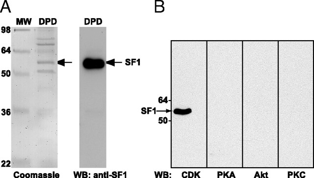
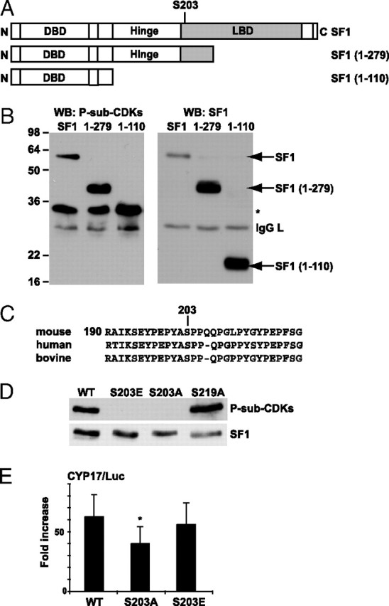
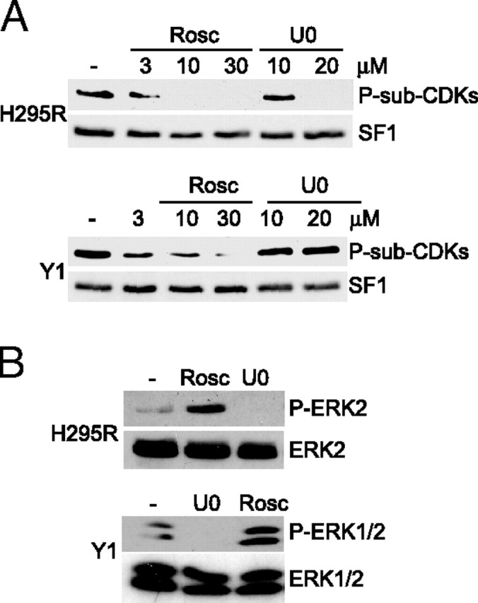
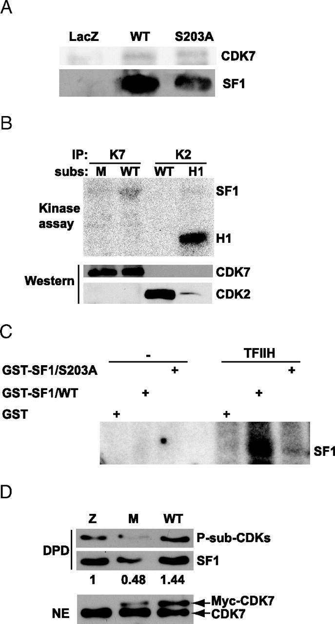
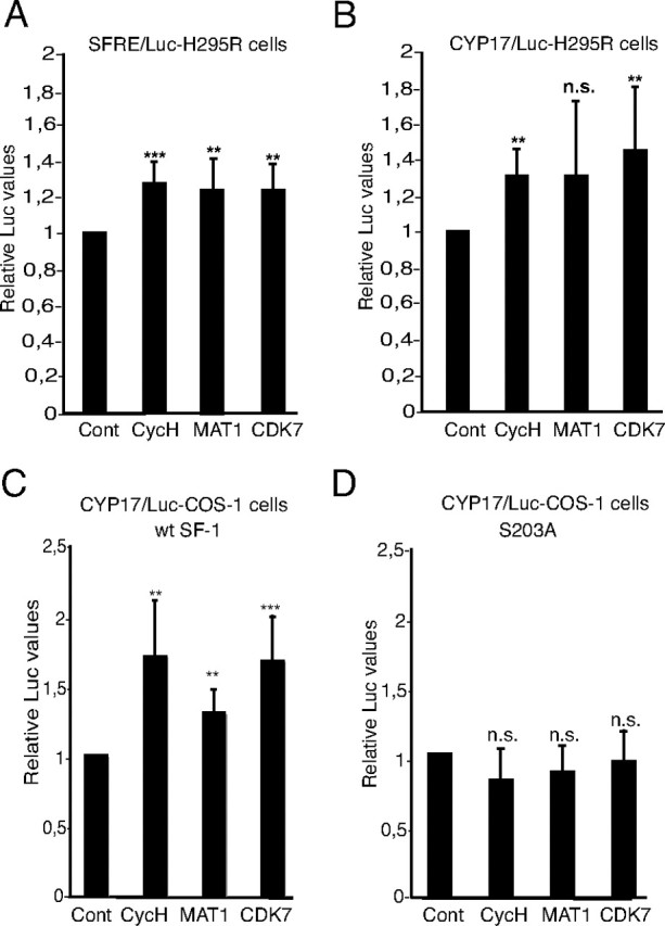
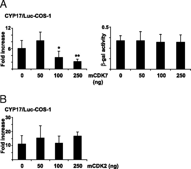
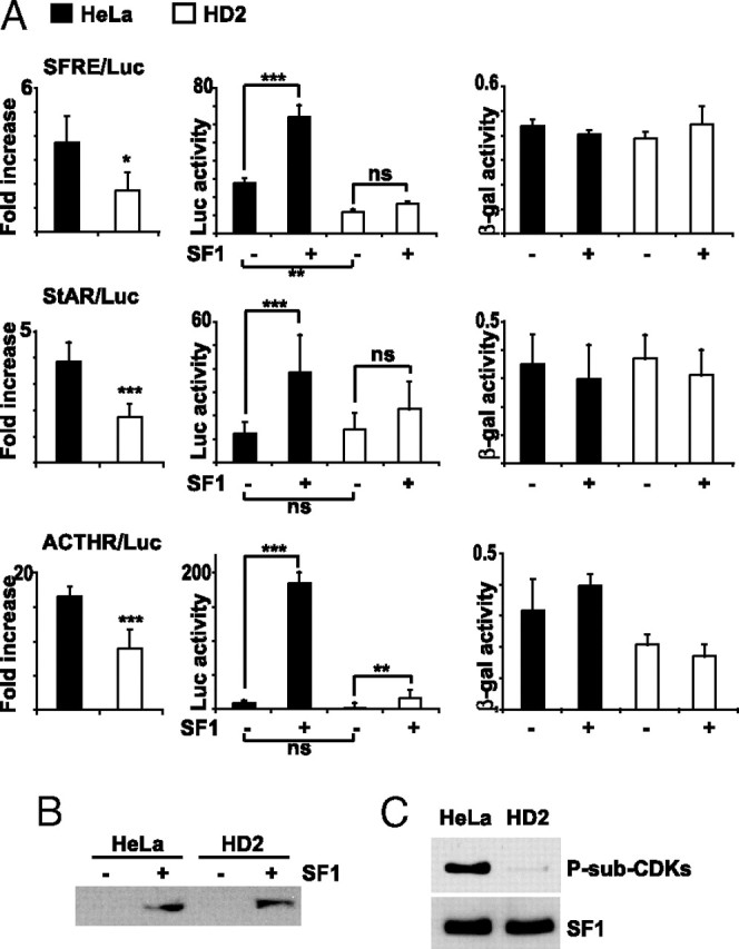
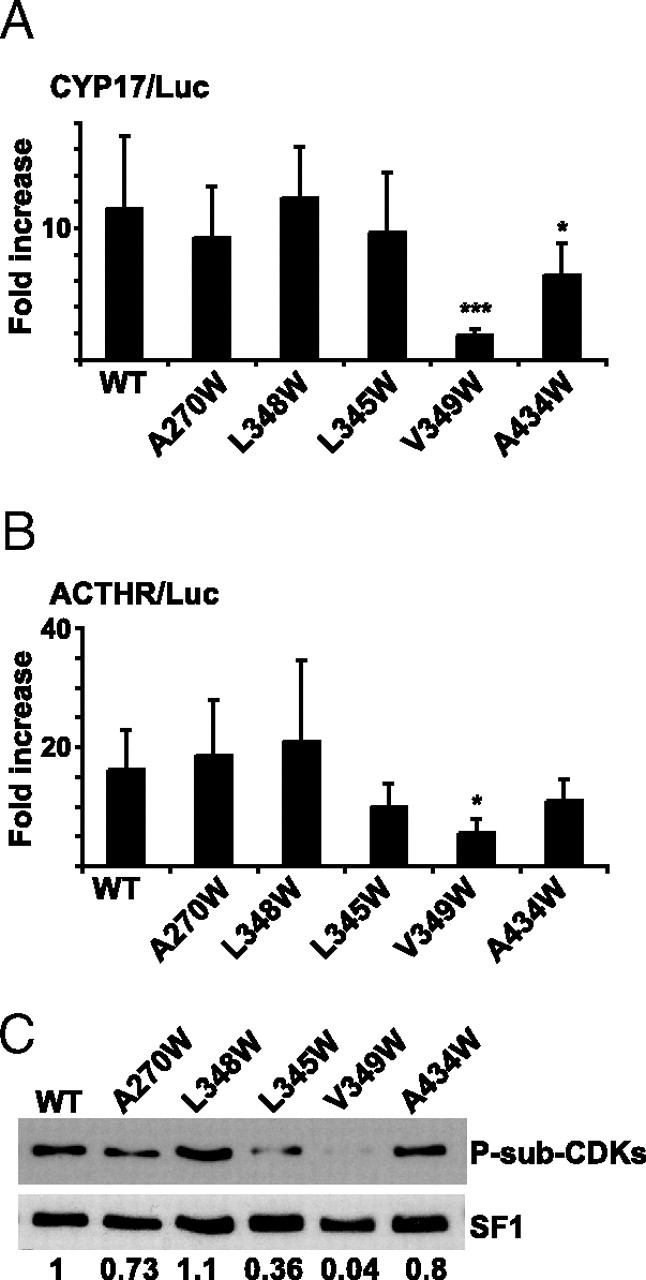
The nuclear receptor steroidogenic factor-1 (SF1) is critical for development and function of steroidogenic tissues. Posttranslational modifications are known to influence the transcriptional capacity of SF1, and it was previously demonstrated that serine 203 is phosphorylated. In this paper we report that serine 203 is phosphorylated by a cyclin-dependent kinase 7 (CDK7)-mediated process. As part of the CDK-activating kinase complex, CDK7 is a component of the basal transcription factor TFIIH, and phosphorylation of SF1 as well as SF1-dependent transcription was clearly reduced in cells carrying a mutation that renders the CDK-activating kinase complex unable to interact with the TFIIH core. Coimmunoprecipitation analyses revealed that SF1 and CDK7 reside in the same complex, and kinase assays demonstrated that immunoprecipitated CDK7 and purified TFIIH phosphorylate SF1 in vitro. The CDK inhibitor roscovitine blocked phosphorylation of SF1, and an inactive form of CDK7 repressed the phosphorylation level and the transactivation capacity of SF1. Structural studies have identified phosphoinositides as potential ligands for SF1. Interestingly, we found that mutations designed to block phospholipid binding dramatically decreased the level of SF1 phosphorylation. Together our results suggest a connection between ligand occupation and phosphorylation and association with the basic transcriptional machinery, indicating an intricate regulation of SF1 transactivation.


Figures

Fig. 1.
Fig. 1.
SF1 Is Phosphorylated by CDKs A, Endogenous SF1 was isolated from H295R nuclear extract (10 mg) by DPD using a biotinylated double-stranded oligonucleotide containing SFREs and streptavidin-conjugated agarose beads. Eluates (DPD) were run on 10% SDS-PAGE and stained with Coomassie blue (left panel) or subjected to Western blotting with an SF1 antibody (right panel). The proteins present in the bands (in the left panel were identified by mass spectrometry (please see main text for details). MW, Molecular weight standard. B, H295R cells were treated with okadaic acid (100 nm) for 1 h, and SF1 was isolated by DPD. Eluates were subjected to Western blotting with the anti-phospho-(Ser and/or Thr) kinase substrate antibodies for CDKs, PKA, Akt, and PKC. WB, Western blot.
Fig. 2.
Fig. 2.
SF1 Is Phosphorylated by CDKs on S203 A, Schematic representation of full-length SF1 and the deletion constructs of SF1 [SF1(1–279), SF1(1–110)] used in this study. The numbering corresponds to the aa sequence. B, COS-1 cells were transfected with Flag-pCMV2/SF1 (SF1), Flag-pCMV2/SF1(1–110) (1–110), and Flag-pCMV2/SF1(1–279) (1–279), and SF1 was immunoprecipitated with the SF1 antibody and subjected to Western blot analyses using the anti-P-S-sub-CDKs antibody and subsequently the SF1 antibody. *, Unknown reactive protein. C, Sequence alignment of human, mouse, and bovine SF1. D, COS-1 cells were transfected with Flag-pCMV2/SF1 (WT) and mutants Flag-pCMV2/SF1(S203A) (S203A), Flag-pCMV2/SF1(S203E) (S203E), or Flag-pCMV2/SF1(S219A) (S219A). SF1 was immunoprecipitated with Flag antibodies, and Western blot analyses were performed using the anti-P-sub-CDKs antibody (upper panel) followed by incubation with SF1 antibodies (lower panel). E, COS-1 cells were transfected with CYP17/Luc (1 μg) together with Flag-pCMV2/SF1 WT, S203A, or S203E (100 ng). The luciferase activity is shown as fold increase and relative to β-gal activity (n = 7–10; *, P < 0.05 as determined by ANOVA, Bonferoni test). DBD, DNA-binding domain; WB, Western blot.
Fig. 3.
Fig. 3.
Inhibition of CDKs Blocks Phosphorylation of SF1 in Adrenocortical Cells A, H295R and Y1 cells were incubated with DMSO (−), increasing concentrations of roscovitine (Rosc; 3, 10, or 30 μm) or U0186 (U0; 10 or 20 μm) for 1 h. SF1 was isolated by DPD, and Western blot analyses were performed using the anti-P-S-sub-CDKs antibody followed by the SF1 antibody. B, H295R and Y1 cells were incubated with DMSO (−), U0186 (U0; 10 μm), or roscovitine (Rosc; 30 μm) for 45 min. Cell extracts were run on SDS-PAGE and blotted with the phospho-ERK1/2 antibody (upper panel) followed by the ERK1/2 antibody (lower panel).
Fig. 4.
Fig. 4.
SF1 Coimmunoprecipitates with CDK7 and Is Phosphorylated by CDK7 and TFIIH A, COS-1 cells were transfected with Flag-pCMV2/SF1 WT or S203A, or the control vector pCMV5/LacZ and immunoprecipitated with the Flag antibody. Eluates were run on a 10% SDS-PAGE and subjected to Western blot analyses using CDK7 (upper panel) and SF1 (lower panel) antibodies. B, CDK7 and CDK2 were immunoprecipitated from H295R cells and incubated with different substrates (subs), GST-SF1(179–431) (WT), GST-SF1(179–431/S203A) (M), or histone H1 (H1) for kinase assays. Membranes were subjected to autoradiography (O/N exposure) followed by Western blot analyses with anti-CDK7 and anti-CDK2. C, GST, GST-SF1(179–431), and GST-SF1(179–431/S203A) were incubated in the absence or presence of TFIIH isolated from HeLa cells. Gels were subjected to autoradiography (1–2 h exposure). D, H295R cells were transfected with control vector pCMV5/LacZ, Myc-CDK7 WT, or the Myc-CDK7-K41R (5 μg). SF1 was then isolated by DPD, and Western blotting was performed with anti-P-Sub-CDK (upper panel) and SF1 (middle panel) antibodies. Nuclear extracts (20 μg) were also subjected to Western blotting with anti-CDK7 (lower panel). Densitometric analyses were performed and are shown as ratios between phosphorylated SF1 and total SF1. The value from control cells was set to 1. IP, Immunoprecipitation; NE, nuclear extract.
Fig. 5.
Fig. 5.
Subunits of the CAK Complex Enhance SF1-Dependent Transcription A, H295R cells were transfected with SFRE/Luc (1 μg) together with expression plasmids encoding cycH (25 ng), MAT1 (10 ng), or CDK7 (10 ng). B, H295R cells were transfected as described in panel A, but with CYP17/Luc instead of SFRE/Luc. C, COS-1 cells were transfected with the CYP17/Luc, and an expression plasmid encoding SF1 (50 ng) together with cycH (50 ng), MAT1 (10 ng), or CDK7 (25 ng) expression plasmids as indicated in the figure. D, COS-1 cells were transfected as described in panel C, but with SF1(S203A) instead of SF1(WT). For all experiments, the luciferase activities in extracts from cells without exogenous expression of cycH, MAT1, or CDK7 were set to 1. Luciferase activities were calculated relative to β-gal activity (from pCMV5/βgal; 100 ng). All transfections were performed in triplicate repeated at least three times, shown as means ± sds. *, P < 0.05, as determined by Student’s t test. Cont, Control; n.s., not significant [P = 0.12 in panel B; P = 0.91 (cycH), 0.83 (MAT1), and 0.97 (CDK7) in panel D].
Fig. 6.
Fig. 6.
Mutated CDK7 Inhibits SF1-Dependent Transactivation COS-1 cells were transfected with CYP17/Luc (1 μg) together with expression plasmids encoding SF1 and a mutated form of CDK7 (CDK7-K41R) (mCDK7; 50–250 ng) (panel A) or CDK2 (mCDK2; 50–250 ng) (panel B). The luciferase activities are given as fold induction relative to the luciferase activities in cells not transfected with Flag-pCMV2/SF1. Luciferase activities were calculated relative to β-gal activity (from pCMV5/βgal; 100 ng). The experiments were repeated at least three times (n ≥11), shown as means ± sds. *, P < 0.05; **, P < 0.01, as determined by one-way ANOVA Bonferoni posttest. The effect of mCDK7 on the pCMV5/βGAL promoter activity is shown in A, right panel.
Fig. 7.
Fig. 7.
Mutation in XPD of TFIIH Decreases SF1-Dependent Transcription and Phosphorylation A, Hela and HD2 cells were transfected with SFRE/Luc, StAR/Luc, or MC2R/Luc (0.5 μg) and pCMV5/LacZ, in the absence or presence of Flag-pCMV2/SF1 (50 ng). The luciferase values are shown as fold increase in the presence of SF1 and relative to the β-gal activity (left panels). The luciferase and the β-gal activities are also shown separately in the middle and right panels, respectively. The experiments were repeated two or three times, shown as means ± sds (SFRE/Luc, n = 5; StAR/Luc, n =12; MC2R/Luc, n = 6). *, P < 0.05; **, P < 0.01; ***, P < 0.001; as determined by unpaired t test (folds) or by ANOVA, Bonferonni (luciferase and β-gal activities). B, Lysates from panel A were incubated with 0.1% SDS and subjected to Western blot analyses with a SF1 antibody to verify equal SF1 expression. C, HeLa and HD2 cells were transfected with Flag-pCMV2/SF1. SF1 was isolated by DPD and Western blotted with anti-P-Sub-CDK (upper panel) and SF1 (lower panel) antibodies. ACTHR, ACTH receptor.
Fig. 8.
Fig. 8.
Mutations in the LBP that Reduce Transcriptional Activity Is Less Phosphorylated by CDKs COS-1 cells were transfected with pCDNA3.1+/SF1 (WT; 50 ng) or LBP mutants (pCDNA3.1Zeo+/SF1-A270W, L348W, L345W, V349W, and A434W; 50 ng) together with the reporter gene construct CYP17/Luc (0.5 μg) (panel A) or MC2R/Luc (panel B). The luciferase activities are presented as fold induction compared with the activity in the absence of SF1 and relatively to the activities from pCMV5/βgal. The experiments were repeated three times, shown as means ± sds (CYP17/Luc, n = 9; *, P < 0.05; ***, P < 0.001; as determined by one-way ANOVA, Bonferoni posttest; MC2R/Luc, n = 6; *, P < 0.05, as determined by nonparametric ANOVA, Dunn posttest). C, COS-1 cells were transfected with SF1 (WT) or LBP mutants (5 μg), and SF1 was isolated by DPD. The isolated proteins were subjected to Western blotting analyses using anti-P-Sub-CDK (upper panel) and SF1 antibodies (lower panel). Densitometric analyses were performed and are shown as ratios between phosphorylated SF1 and total SF1. ACTHR, ACTH receptor.

Similar articles

Cited by

References

    1. Parker KL, Rice DA, Lala DS, Ikeda Y, Luo X, Wong M, Bakke M, Zhao L, Frigeri C, Hanley NA, Stallings N, Schimmer BP 2002. Steroidogenic factor 1: an essential mediator of endocrine development. Recent Prog Horm Res 57:19–36 - PubMed
    1. Wang W, Zhang C, Marimuthu A, Krupka HI, Tabrizizad M, Shelloe R, Mehra U, Eng K, Nguyen H, Settachatgul C, Powell B, Milburn MV, West BL 2005. The crystal structures of human steroidogenic factor-1 and liver receptor homologue-1. Proc Natl Acad Sci USA 102:7505–7510 - PMC - PubMed
    1. Krylova IN, Sablin EP, Moore J, Xu RX, Waitt GM, MacKay JA, Juzumiene D, Bynum JM, Madauss K, Montana V, Lebedeva L, Suzawa M, Williams JD, Williams SP, Guy RK, Thornton JW, Fletterick RJ, Willson TM, Ingraham HA 2005. Structural analyses reveal phosphatidyl inositols as ligands for the NR5 orphan receptors SF-1 and LRH-1. Cell 120:343–355 - PubMed
    1. Li Y, Choi M, Cavey G, Daugherty J, Suino K, Kovach A, Bingham NC, Kliewer SA, Xu HE 2005. Crystallographic identification and functional characterization of phospholipids as ligands for the orphan nuclear receptor steroidogenic factor-1. Mol Cell 17:491–502 - PubMed
    1. Urs AN, Dammer E, Sewer MB 2006. Sphingosine regulates the transcription of CYP17 by binding to steroidogenic factor-1. Endocrinology 147:5249–5258 - PubMed

Publication types

MeSH terms