. 2007 Oct 9;104(41):16080-5.
doi: 10.1073/pnas.0702451104. Epub 2007 Sep 27.

Disease-associated mutant alpha-actinin-4 reveals a mechanism for regulating its F-actin-binding affinity

Affiliations

Disease-associated mutant alpha-actinin-4 reveals a mechanism for regulating its F-actin-binding affinity

Astrid Weins et al. Proc Natl Acad Sci U S A. .

Abstract

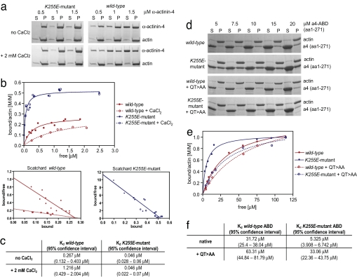
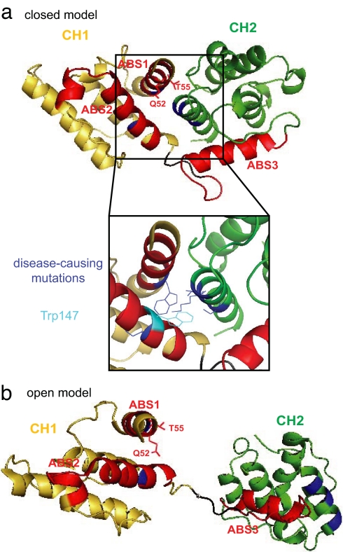
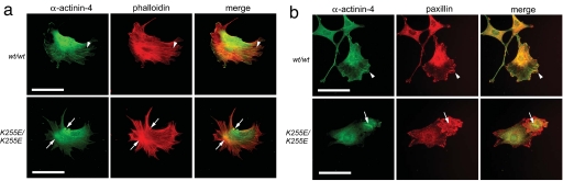
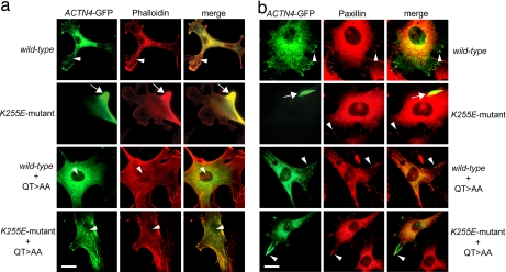
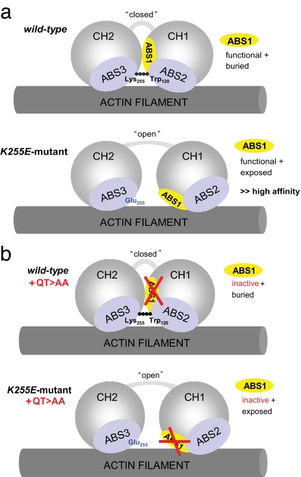
Alpha-actinin-4 is a widely expressed protein that employs an actin-binding site with two calponin homology domains to crosslink actin filaments (F-actin) in a Ca(2+)-sensitive manner in vitro. An inherited, late-onset form of kidney failure is caused by point mutations in the alpha-actinin-4 actin-binding domain. Here we show that alpha-actinin-4/F-actin aggregates, observed in vivo in podocytes of humans and mice with disease, likely form as a direct result of the increased actin-binding affinity of the protein. We document that exposure of a buried actin-binding site 1 in mutant alpha-actinin-4 causes an increase in its actin-binding affinity, abolishes its Ca(2+) regulation in vitro, and diverts its normal localization from actin stress fibers and focal adhesions in vivo. Inactivation of this buried actin-binding site returns the affinity of the mutant to that of the WT protein and abolishes aggregate formation in cells. In vitro, actin filaments crosslinked by the mutant alpha-actinin-4 exhibit profound changes of structural and biomechanical properties compared with WT alpha-actinin-4. On a molecular level, our findings elucidate the physiological importance of a dynamic interaction of alpha-actinin with F-actin in podocytes in vivo. We propose that a conformational change with full exposure of actin-binding site 1 could function as a switch mechanism to regulate the actin-binding affinity of alpha-actinin and possibly other calponin homology domain proteins under physiological conditions.


Conflict of interest statement

The authors declare no conflict of interest.

Figures

Fig. 1.
Fig. 1.
K255E mutant α-actinin-4 shows higher F-actin-binding affinity as the WT protein and is resistant to Ca2+ regulation. (a) Representative images of Coomassie-blue-stained polyacrylamide gels at three molar ratios. Increasing amounts of K255E mutant and WT α-actinin-4 without (upper gels) or with (lower gels) the addition of 2 mM CaCl2 to reaction buffer were polymerized with 5 μM G-actin and centrifuged at 100,000 × g. Equal amounts of supernatants and pellets were subjected to SDS/PAGE. α-Actinin-4 alone did not pellet at this speed (data not shown). (b) K255E mutant α-actinin-4 shows increased binding affinity to F-actin, a higher binding stoichiometry, and is insensitive to Ca2+. Saturation binding curves and Scatchard plots were generated by nonlinear regression from quantified results of Coomassie-blue-stained polyacrylamide gels. (c) The table shows respective Kd values and 95% confidence intervals. Inactivation of ABS1 by introducing the QT>AA mutation into the K255E mutant α-actinin-4 ABD (a4-ABD) returns its F-actin-binding affinity to that of the WT protein. (d) Representative images taken from Coomassie-blue-stained polyacrylamide gels. Increasing amounts (5–120 μM) of purified α-actinin-4 ABDs were polymerized with 5 μM G-actin and centrifuged at 100,000 × g, and equal amounts of supernatants and pellets were subjected to SDS/PAGE. (e) K255E mutant α-actinin-4 ABD shows higher binding affinity to F-actin, whereas K255E mutant α-actinin-4 ABD containing the ABS1 mutation QT>AA shows similar F-actin-binding affinity as the WT protein. The F-actin-binding affinity of WT α-actinin-4 ABD containing the QT>AA mutation was only minimally affected. (f) The table shows respective Kd values and 95% confidence intervals.
Fig. 2.
Fig. 2.
Structural models of the ABD of α-actinin based on two previously published atomic structures (15, 17). (a) This model is consistent with a closed conformation of the CH domains. CH1 is shown in yellow, CH2 in green. ABS1 to -3 are shown in red; side chains of Q52 and T55, mutated to alanines in the QT>AA mutant, are shown to demonstrate their inward-facing position within the model. The enlarged view of the CH1–CH2 interface demonstrates that amino acids changed by the five disease-associated mutations (W59R, I149del, K255E, T259I, and S262P), depicted in blue, all localize to the CH1–CH2 interface region. In addition, Lys-255, mutated to Glu in the K255E mutant, interacts with Trp-147 (shown in cyan) of CH1 to form a hinge-like connection that supposedly functions as a key connection that keeps the CH domains in a closed conformation. (b) This model shows an open conformation of the CH domains. Here, the two CH domains are widely separated, thereby fully exposing ABS1, including Q52 and T55. The interface regions, bearing the disease-associated mutations, are pointing in opposite directions, breaking apart all atom-to-atom connections between CH1 and CH2.
Fig. 3.
Fig. 3.
K255E mutant α-actinin-4 crosslinks F-actin into architecturally distinct structures. (a) Transmission electron microscopy of actin filaments alone and crosslinked by WT or K255E mutant α-actinin-4 in vitro. WT α-actinin-4 crosslinks actin filaments into thick parallel bundles with defined spacing. Mutant α-actinin-4 induces the formation of a disordered and entangled network of thin filament bundles. (Scale bars, 200 nm.) (b) Viscometry assay of actin in the presence of WT or K255E mutant α-actinin-4. Decreasing α-actinin-4/actin molar ratios were used for gel point assays and are expressed as a log scale. Solid-to-gel transition is shown by the ∞ sign. The gel point concentration of WT α-actinin-4 was determined at 1:100, whereas the gel point of the K255E mutant was found at 1:850. (c) Determination of actin gel points in the presence of filamin A and WT and K255E mutant α-actinin-4 as different actin filament lengths. The gel point concentrations are shown as a molar ratio of crosslinker to actin and at four different gelsolin concentrations. WT α-actinin-4 can only induce actin gelation with long filaments. With decreasing filament length, a higher concentration of K255E mutant α-actinin-4 than filamin A is required to induce gelation.
Fig. 4.
Fig. 4.
Endogenous K255E mutant α-actinin-4 induces cytoplasmic F-actin aggregates in immortalized lung fibroblasts and is absent from peripheral stress fibers and focal adhesions. (a) Representative images of fibroblasts isolated from Actn4WT/WT and Actn4K255E/K255E mice. Cells were immunostained with anti-α-actinin-4 (green) and rhodamine-phalloidin (red). Images were merged; areas of colocalization appear yellow. Arrowheads point to expression of α-actinin-4 along peripheral actin stress fibers in WT/WT cells. In K255E/K255E cells, peripheral expression of α-actinin-4 is notably diminished, and arrows point to F-actin aggregates. (b) Representative images of fibroblasts isolated from Actn4WT/WT and Actn4K255E/K255E mice. Cells were immunostained with anti-α-actinin-4 (green) and anti-paxillin (red), and images were merged. Arrowheads point to colocalization of α-actinin-4 with paxillin at focal adhesions in WT/WT cells. In K255E/K255E cells, α-actinin-4 is absent from focal adhesions and the cell periphery and forms cytoplasmic aggregates (arrows). (Scale bars, 25 μm.)
Fig. 5.
Fig. 5.
Actn4-deficient fibroblasts expressing WT or K255E mutant ACTN4–GFP containing the QT>AA mutation exhibit normal distribution of α-actinin-4–GFP. (a) Actn4-deficient fibroblasts were transfected with WT or K255E mutant ACTN4–GFP (green) without and with the QT>AA mutation and were stained with rhodamine–phalloidin (red). Images were merged; areas of colocalization appear yellow. Arrowheads point to normal distribution of α-actinin-4–GFP along actin stress fibers in cells expressing WT, WT+QT>AA, and K255E mutant+QT>AA ACTN4 constructs. In contrast, K255E mutant-expressing cells show α-actinin-4 staining associated exclusively with a large peripheral aggregate that also exhibits strong phalloidin staining, whereas F-actin stress fibers are absent. (b) Actn4-deficient fibroblasts were transfected with the various ACTN4–GFP constructs (green) and coimmunostained with an antibody against paxillin, a focal adhesion marker (red). All transfected cells exhibit paxillin staining at focal adhesion sites (arrowheads). Cells expressing WT, WT+QT>AA, or K255E mutant+QT>AA ACTN4–GFP showed normal peripheral expression of α-actinin-4 at focal adhesions that colocalized with paxillin staining. In contrast, K255E mutant-expressing cells show α-actinin-4 staining exclusively in a large aggregate (arrows), and α-actinin-4–GFP is absent from focal adhesions in these cells. (Scale bars, 25 μm.)
Fig. 6.
Fig. 6.
Schematic illustration of the binding of the CH domains of WT and K255E mutant α-actinin-4 in open or closed conformation to an actin filament involving ABS1 to -3. (a) In the WT protein, the CH domains can adopt a closed conformation in which ABS1 (yellow) is buried between the CH domains. ABS2 and -3 (light blue) mainly account for F-actin binding in this conformation. The Lys-255–Trp-147 hinge-like connection contributes to keep the CH domains in a closed conformation. In the mutant protein, because of a Lys>Glu amino acid exchange at amino acid 255 (mutation shown in blue), the connection with Trp-147 is lost and the two CH domains adopt an “open” conformation. ABS1 is exposed and extends the actin-binding surface together with ABS2 and -3. (b) Inactivation of ABS1 (illustrated by the red crossed bars) by the QT>AA mutation does not affect actin binding of the WT protein because ABS1 does not contribute to actin binding in the closed conformation. In the K255E mutant, however, in which ABS1 is exposed because of the open conformation of the CH domains, ABS1 inactivation returns the actin-binding affinity back to that of the WT protein.

Similar articles

Cited by

References

    1. Maruyama K, Ebashi S. J Biochem (Tokyo) 1965;58:13–19. - PubMed
    1. Mimura N, Asano A. Nature. 1978;272:273–276. - PubMed
    1. Pelletier O, Pokidysheva E, Hirst LS, Bouxsein N, Li Y, Safinya CR. Phys Rev Lett. 2003;91:148102. - PubMed
    1. Wachsstock DH, Schwarz WH, Pollard TD. Biophys J. 1994;66:801–809. - PMC - PubMed
    1. Witke W, Schleicher M, Noegel AA. Cell. 1992;68:53–62. - PubMed

Publication types

MeSH terms