. 2007 Oct 15;21(20):2593-606.
doi: 10.1101/gad.1592107. Epub 2007 Sep 27.

The requirement for Phr1 in CNS axon tract formation reveals the corticostriatal boundary as a choice point for cortical axons

Affiliations

The requirement for Phr1 in CNS axon tract formation reveals the corticostriatal boundary as a choice point for cortical axons

A Joseph Bloom et al. Genes Dev. .

Abstract

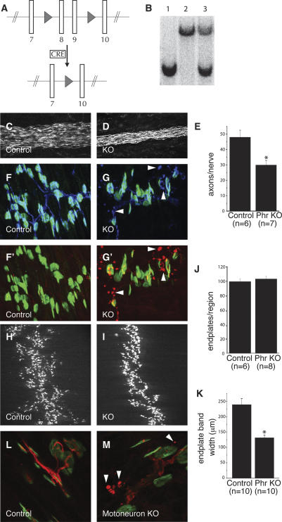
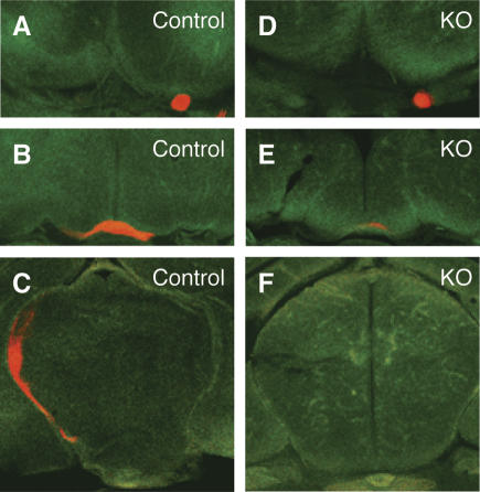
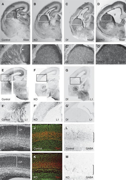
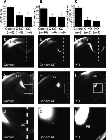
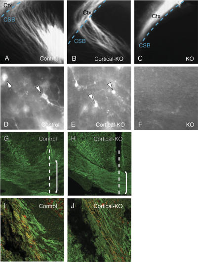
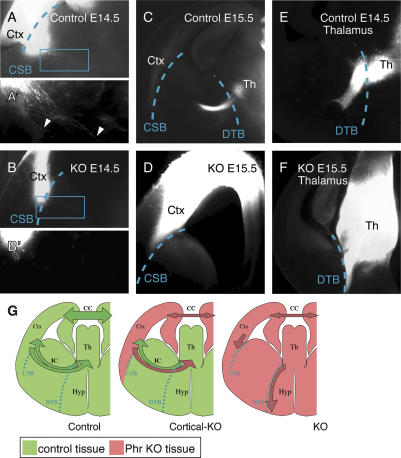
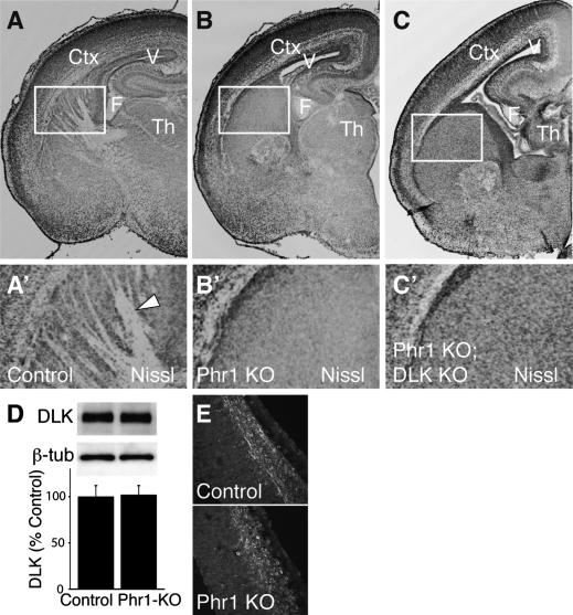
Phr1 is the single well-conserved murine ortholog of the invertebrate ubiquitin ligase genes highwire (in Drosophila) and rpm-1 (in Caenorhabditis elegans). The function and mechanism of action of highwire and rpm-1 are similar--both cell-autonomously regulate synaptogenesis by down-regulating the ortholog of the mitogen-activated protein kinase kinase kinase dual leucine zipper kinase (MAPKKK DLK). Here, using a targeted conditional mutant, we demonstrate that Phr1 also plays essential roles in mammalian neural development. As in invertebrates, Phr1 functions cell-autonomously to sculpt motor nerve terminals. In addition, Phr1 plays essential roles in the formation of major CNS axon tracts including those of the internal capsule, in part via cell-nonautonomous mechanisms, and these results reveal a choice point for cortical axons at the corticostriatal boundary. Furthermore, whereas the neurite morphology phenotypes of highwire and rpm-1 are suppressed by loss of DLK in flies and worms, Phr1-dependent CNS defects persist in Phr1, DLK double mutants. Thus, in the mammalian nervous system Phr1 is required for formation of major CNS axon tracts via a mechanism that is both cell-nonautonomous and independent of DLK.


Figures

Figure 1.
Figure 1.
Phr1 functions in the motoneuron to restrain presynaptic sprouting. (A) Schematic of the targeted Phr1 mutant allele. Two LoxP sites (triangles) were introduced by homologous recombination into the Phr1 locus flanking the eighth and ninth translated exons to create the “floxed” allele. In the presence of Cre, the two LoxP sites recombine and excise the intervening genomic DNA to become the constitutive knockout allele. (B) Southern analysis demonstrates the presence of the targeted chromosome in the Phr1 knockout homozygote (lane 1), the wild-type chromosome in a wild-type littermate (lane 2), and both chromosomes in a heterozygote (lane 3). Phrenic nerves stained with neurofilament are shown at E18.5 from control (C) and Phr1 constitutive knockout (KO) (D) embryos. Quantification of the mean number of axons in the phrenic nerve is shown in E. NMJs from E18.5 control (F,F′) and Phr1 constitutive knockout (G,G′) diaphragms are shown stained for acetylcholine receptors with bungarotoxin (green), synaptophysin (blue), and neurofilament (red). In the Phr1 constitutive knockout, examples of extrasynaptic sprouts containing neurofilament and synaptophysin are highlighted with arrowheads. The endplate band at E18.5 stained for acetylcholine receptors with bungarotoxin is shown for control (H) and Phr1 constitutive knockout (I) diaphragms. Quantification of the mean number of endplates within a defined muscle region as a percentage of wild type is shown in J. Quantification of the mean endplate band width is shown in K. NMJs from P3 control (L) (Hb9-Cre; +) and Phr1 motoneuron knockout (M) (Hb9-Cre; Phr1flox/Phr1flox) diaphragms are shown stained for acetylcholine receptors with bungarotoxin (green) and neurofilament (red). In the Phr1 motoneuron knockout, examples of extrasynaptic sprouts containing neurofilament are highlighted with arrowheads. (*) P < 0.01. Data are presented as mean ± SEM.
Figure 2.
Figure 2.
Phr1 mutants lack retinal innervation of the thalamus. Green Nissl-stained serial coronal sections from P0 heads labeled with DiI (red) in the retina. Labeled optic nerves (A,D), optic chiasm (B,E), and thalamus (C,F) from control and Phr1 constitutive knockouts. Axon labeling is greatly reduced at the knockout chiasm and is absent in the knockout thalamus.
Figure 3.
Figure 3.
Phr1 constitutive knockout displays severe defects of gross morphology and axon fiber tracts in the CNS. Nissl-stained coronal sections from one side of E18.5 brains of control (A,A′), Phr1 constitutive knockout (KO) (B,B′), Phr1 deficiency mutants (15DttMb/15DttMb) (C,C′), and transheterozygotes for the constitutive allele and deficiency allele (D,D′). Nissl stains cell bodies and shows white matter tracts in relief. In the Phr1 knockout and Phr1 deficiency mutant (Df), lateral ventricles are increased in size, the hippocampus is reduced, and axon tracts running through the internal capsule are absent. Cortex (Ctx), lateral ventricle (V), fimbria (F), and thalamus (Th) are labeled. The boxed region is shown in A′, B′, C′, and D′ and demonstrates the presence of white matter tracts (arrowhead) in the internal capsule from the control (A′) but not from the Phr1 knockout and Phr1 deficiency mutant (B′,C′,D′). Staining for L1, a marker of major fiber tracts including the internal capsule, is shown in coronal sections from one side of E18.5 brains of control (E,E′), Phr1 constitutive knockout (KO) (F,F′), and Phr1 deficiency mutants (Df) (G,G′). The boxed regions shown in E′, F′, and G′ demonstrate the presence of L1-positive axon tracts (arrowhead) in the internal capsule from the control (E′) but not of the Phr1 knockout (F′) or Phr1 deficiency mutant (G′). Coronal sections of Nissl-stained control (H) and Phr1 constitutive knockout (KO) (I) E18.5 cerebral cortex highlight the cortical plate (CP) whose thickness (indicated by the bracket) as a percentage of the total cortical thickness is unchanged in the mutant. Coronal sections of Nissl-stained (green) and Tbr1-stained (red) control (J) and Phr1 constitutive knockout (KO) (K) E18.5 cerebral cortex highlight that Tbr1+ neurons have migrated into the cortex and differentiated in similar positions in mutant and controls. Coronal sections of GABA-stained control (L) and Phr1 constitutive knockout (KO) (M) E18.5 cerebral cortex show GABAergic cell bodies (arrowheads) in both the control and mutant, while highlighting the more prominent GABAergic neurite staining (bracketed regions) in the control.
Figure 4.
Figure 4.
Corpus callosum defects in the Phr1 mutant are region specific. Quantification of corpus callosum size as measured from brain sections of E18.5 control, cerebral cortex-specific Phr1 knockout (C-KO, Emx1-Cre;Phr1flox/Phr1flox), and constitutive Phr1 knockout (KO) shows the anterior–posterior depth (A), dorsal–ventral thickness (B), and cross-sectional area (C). In A, the asterisk indicates P < 0.01, in B, the asterisk indicates P < 0.005, and in C, the asterisk indicates P < 0.0005 compared with control, demonstrating that Phr1 is required for normal corpus callosum formation. The size of the corpus callosum is not different between the cerebral cortex-specific and constitutive Phr1 alleles, demonstrating that Emx1-Cre effectively deletes Phr1 in the projection neurons contributing to the corpus callosum. DiI labeling of cortical axons is shown in E18.5 coronal sections in D–L. DiI placed in the somatosensory cortex (Ctx) of control (D), cerebral cortex-specific Phr1 knockouts (Cortical-KO) (E), and constitutive Phr1 knockouts (KO) (F) labels axons that cross the midline (dashed line) via the corpus callosum. Asterisk indicates dye placement in the cortex. DiI placed in the visual cortex of control (G,J), cerebral cortex-specific Phr1 knockouts (Cortical-KO) (H,K), and constitutive Phr1 knockouts (KO) (I,L) labels axons that cross the midline (dashed line) in the control, but not in the cerebral cortex-specific or constitutive Phr1 knockout. The boxed region in G–I is magnified in J–L and highlights that axons from the visual cortex cross the midline in the control, but fail to cross and instead accumulate in “Probst bundles” in both the cerebral cortex-specific and constitutive Phr1 knockout. Data are presented as mean ± SEM.
Figure 5.
Figure 5.
The cortical axon outgrowth defect of Phr1 mutants is cell-nonautonomous. DiI labeling is shown in E18.5 coronal sections in A–F. DiI placed in the somatosensory cortex (Ctx) of control (A) and cerebral cortex-specific Phr1 knockouts (Cortical-KO) (B) labels axons that cross the corticostriatal boundary (CSB, dashed line) and traverse the internal capsule. (C) When DiI is placed in the somatosensory cortex of the constitutive Phr1 knockouts (KO), axons remain in the cortex. When DiI is placed in the visual cortex of control (D) or the cerebral cortex-specific Phr1 knockout (E), cell bodies in the dorsal thalamus are labeled, demonstrating that thalamocortical projections have reached the cortex in these genotypes. (F) DiI placement in the visual cortex of constitutive Phr1 knockouts (KO) fails to label retrogradely thalamic neurons. To visualize selectively Cre-expressing cortical neurons, Thy1-stop-YFP transgene mice were mated to produce control (Emx1-Cre, Thy1-stop-YFP) (G,I) and the cerebral cortex-specific Phr1 knockouts (Cortical-KO: Emx1-Cre, Phr1flox/Phr1flox, Thy1-stop-YFP) (H,J). YFP-positive axons cross the midline (dashed line) and contribute to the corpus callosum (bracketed region) in both the control (G) and cortical knockout (H). Note that the corpus callosum is thinner in the cortical knockout as expected for a Phr1 knockout. YFP-positive axons also contribute to the internal capsule in both the control (I) and cortical knockout (J). In I and J, Cre-expressing cortical axons in the internal capsule are labeled with YFP (green), while thalamocortical axons in the internal capsule are labeled with calretinin (red). The presence of cortical axons in the internal capsule of the cerebral cortex-specific Phr1 knockout, but not the constitutive Phr1 knockout, demonstrates that this cortical axon defect is non-cell-autonomous.
Figure 6.
Figure 6.
Axonal projections in the Phr1 mutant reveal guidance cues at the corticostriatal boundary. (A) DiI placed in the somatosensory cortex (Ctx) of controls at E14.5 labels axons that cross the corticostriatal boundary (CSB, dashed line). The boxed region is magnified in A′ and highlights axons (arrowheads) extending deep into the subcortical telencephalon. (B) DiI placed in the somatosensory cortex (Ctx) of constitutive Phr1 knockouts (KO) at E14.5 labels axons that stop at the corticostriatal boundary (CSB, dashed line). The boxed region is magnified in B′and reveals the absence of axons extending into the subcortical telencephalon. (C) At E15.5, DiI placed in the somatosensory cortex (Ctx) of controls labels axons that have crossed the diencephalon–telencephalon boundary (DTB, dashed line) and entered the thalamus (Th). (D) At E15.5, DiI placed in the somatosensory cortex (Ctx) of constitutive Phr1 knockouts (KO) labels axons that remain stalled at the corticostriatal boundary. These results demonstrate that cortical axons in the Phr1 mutant do not cross the corticostriatal boundary. (E) DiI placed in the thalamus (Th) of controls at E14.5 labels thalamic axons that cross the diencephalon–telencephalon boundary (DTB, dashed line) and enter the telencephalon. (F) DiI placed in the thalamus (Th) of constitutive Phr1 knockouts (KO) at E15.5 labels thalamic axons that fail to cross the diencephalon–telencephalon boundary (DTB, dashed line), but instead course ventrally along the boundary between telencephalon and hypothalamus. (G) A schematic summarizes the results from control, cerebral cortex-specific Phr1 knockouts (Cortical-KO), and constitutive Phr1 knockouts (KO) presented in Figures 5 and 6, where wild-type tissue is green and Phr1 mutant tissue is pink. Labeled structures include the cerebral cortex (Ctx), thalamus (Th), hypothalamus (Hyp), corpus callosum (CC), internal capsule (IC), corticostriatal boundary (CSB), and diencephalon–telencephalon boundary (DTB). The schematic highlights that, in the constitutive Phr1 knockout, (1) the cortical defect is non-cell-autonomous and (2) the corticofugal and thalamocortical axons fail to cross the corticostriatal boundary (CSB) and diencephalon–telencephalon boundary (DTB), respectively, and thus both avoid the subcortical telencephalon.
Figure 7.
Figure 7.
CNS defects persist in Phr1, DLK double mutants. Nissl-stained coronal sections from one side of E18.5 brains of control (A,A′), Phr1 constitutive knockout (Phr1 KO) (B,B′, and Phr1; DLK double knockouts (Phr1 KO; DLK KO) (C,C′). In the Phr1 knockout and Phr1, DLK double knockout, lateral ventricles are increased in size and axon tracts running through the internal capsule are lacking. Cortex (Ctx), lateral ventricle (V), and fimbria (F) are labeled. The boxed regions shown in A′, B′, and C′ demonstrate the presence of white matter tracts (arrowhead) in the internal capsule from the control (A′) but not from the Phr1 knockout (B′) or Phr1, DLK double knockout (C′). These data demonstrate that the major CNS defects in the Phr1 knockout are not suppressed by mutating DLK. In D, Western analysis is shown for DLK and β-tubulin (β-tub) from heads of conditional Phr1 knockout (Phr1-KO) and matching controls. Histogram shows DLK levels relative to β-tubulin loading controls and normalized to control levels. There are no significant differences in the level of DLK between the constitutive Phr1 knockout and control (n = 4 for each genotype). (E) Immunohistochemical localization of DLK in cortical sections of E13.5 cerebral cortex from a constitutive Phr1 knockout (Phr1 KO) and matching control demonstrates no apparent difference in levels or localization of DLK protein. Data are presented as mean ± SEM.

References

    1. Bagri A., Marin O., Plump A.S., Mak J., Pleasure S.J., Rubenstein J.L., Tessier-Lavigne M., Marin O., Plump A.S., Mak J., Pleasure S.J., Rubenstein J.L., Tessier-Lavigne M., Plump A.S., Mak J., Pleasure S.J., Rubenstein J.L., Tessier-Lavigne M., Mak J., Pleasure S.J., Rubenstein J.L., Tessier-Lavigne M., Pleasure S.J., Rubenstein J.L., Tessier-Lavigne M., Rubenstein J.L., Tessier-Lavigne M., Tessier-Lavigne M. Slit proteins prevent midline crossing and determine the dorsoventral position of major axonal pathways in the mammalian forebrain. Neuron. 2002;33:233–248. - PubMed
    1. Bareyre F.M., Kerschensteiner M., Misgeld T., Sanes J.R., Kerschensteiner M., Misgeld T., Sanes J.R., Misgeld T., Sanes J.R., Sanes J.R. Transgenic labeling of the corticospinal tract for monitoring axonal responses to spinal cord injury. Nat. Med. 2005;11:1355–1360. - PubMed
    1. Burgess R.W., Peterson K.A., Johnson M.J., Roix J.J., Welsh I.C., O’Brien T.P., Peterson K.A., Johnson M.J., Roix J.J., Welsh I.C., O’Brien T.P., Johnson M.J., Roix J.J., Welsh I.C., O’Brien T.P., Roix J.J., Welsh I.C., O’Brien T.P., Welsh I.C., O’Brien T.P., O’Brien T.P. Evidence for a conserved function in synapse formation reveals Phr11 as a candidate gene for respiratory failure in newborn mice. Mol. Cell. Biol. 2004;24:1096–1105. - PMC - PubMed
    1. Carney R.S., Alfonso T.B., Cohen D., Dai H., Nery S., Stocia B., Slotkin J., Bregman B.S., Fishell G., Corbin J.G., Alfonso T.B., Cohen D., Dai H., Nery S., Stocia B., Slotkin J., Bregman B.S., Fishell G., Corbin J.G., Cohen D., Dai H., Nery S., Stocia B., Slotkin J., Bregman B.S., Fishell G., Corbin J.G., Dai H., Nery S., Stocia B., Slotkin J., Bregman B.S., Fishell G., Corbin J.G., Nery S., Stocia B., Slotkin J., Bregman B.S., Fishell G., Corbin J.G., Stocia B., Slotkin J., Bregman B.S., Fishell G., Corbin J.G., Slotkin J., Bregman B.S., Fishell G., Corbin J.G., Bregman B.S., Fishell G., Corbin J.G., Fishell G., Corbin J.G., Corbin J.G. Cell migration along the lateral cortical stream to the developing basal telencephalic limbic system. J. Neurosci. 2006;26:11562–11574. - PMC - PubMed
    1. Chapouton P., Schuurmans C., Guillemot F., Gotz M., Schuurmans C., Guillemot F., Gotz M., Guillemot F., Gotz M., Gotz M. The transcription factor neurogenin 2 restricts cell migration from the cortex to the striatum. Development. 2001;128:5149–5159. - PubMed

Publication types

MeSH terms

LinkOut - more resources