Review
. 2007 Oct;13(5):532-45.
doi: 10.1177/1073858407301369.

The prefrontal cortex and flexible behavior

Affiliations
Review

The prefrontal cortex and flexible behavior

Helen Barbas et al. Neuroscientist. 2007 Oct.

Abstract

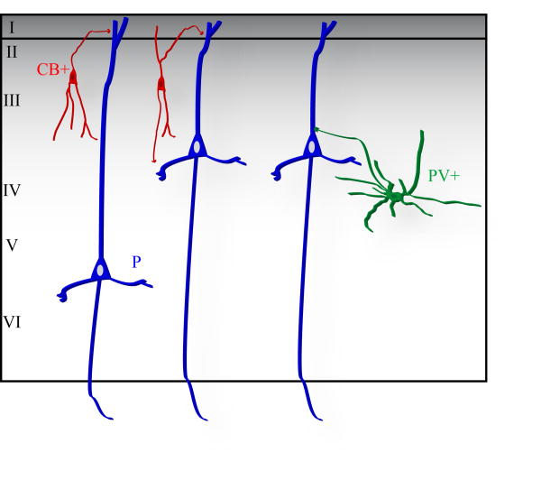
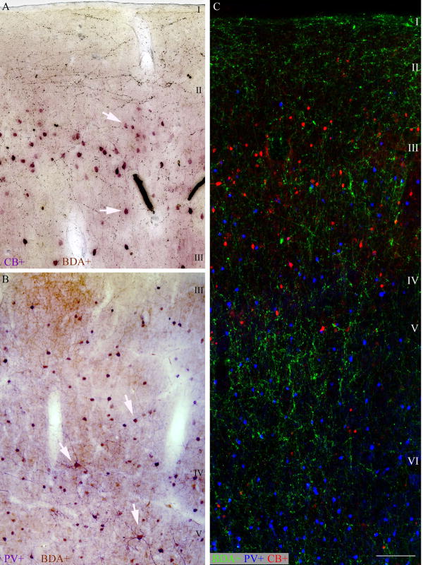
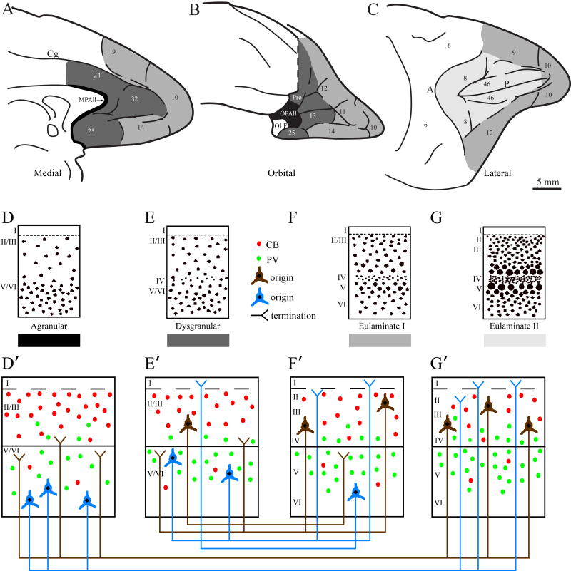
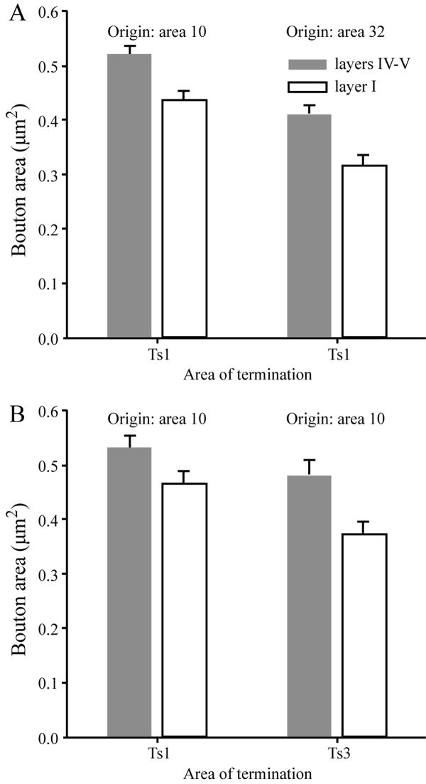
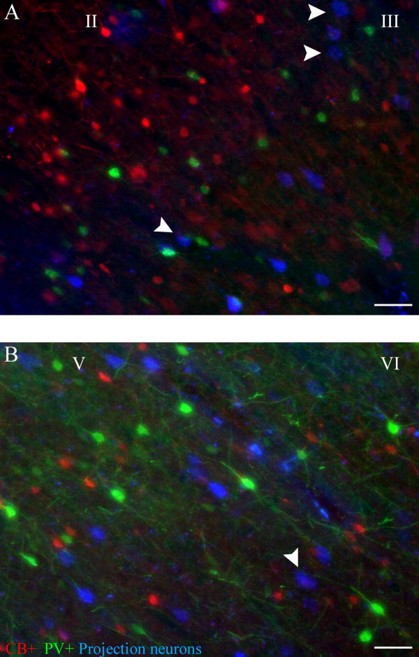
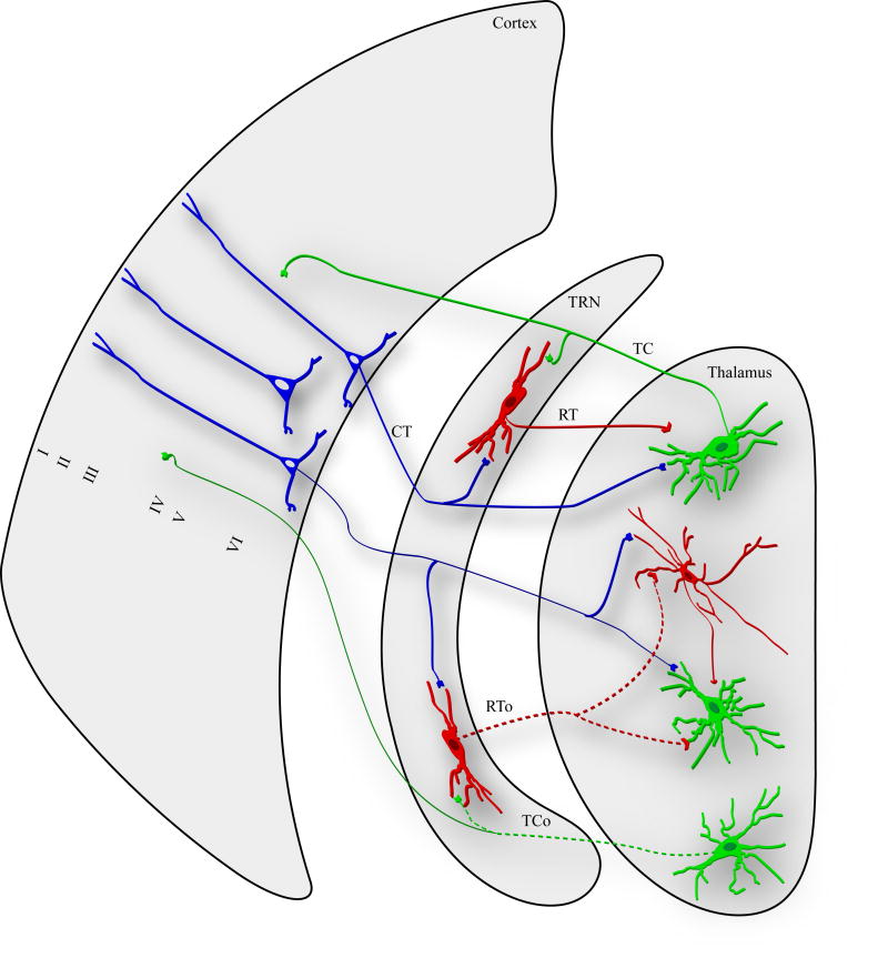
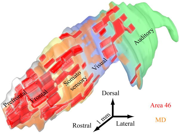
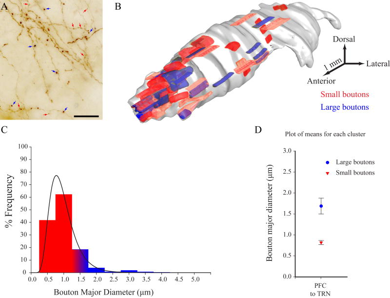
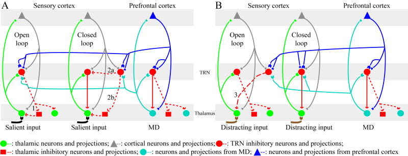
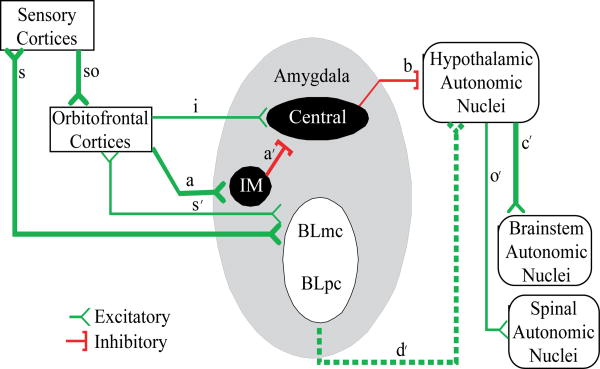
The prefrontal cortex in primates guides behavior by selecting relevant stimuli for the task at hand, mediated through excitatory bidirectional pathways with structures associated with sensory processing, memory, and emotions. The prefrontal cortex also has a key role in suppressing irrelevant stimuli through a mechanism that is not well understood. Recent findings indicate that prefrontal pathways interface with laminar-specific neurochemical classes of inhibitory neurons in sensory cortices, terminate extensively in the frontal and sensory sectors of the inhibitory thalamic reticular nucleus, and target the inhibitory intercalated masses of the amygdala. Circuit-based models suggest that prefrontal pathways can select relevant signals and efficiently suppress distractors, in processes that are disrupted in schizophrenia and in other disorders affecting prefrontal cortices.


Figures

Figure 1
Figure 1
Laminar distribution and pattern of innervation of two neurochemical classes of inhibitory neurons in the cortex. Calbindin neurons (CB, red) are preferentially found in the superficial cortical layers and innervate the distal dendrites of pyramidal neurons (blue) from other layers. Parvalbumin inhibitory neurons (PV, green) are preferentially found in the middle layers of the cortex, and innervate the proximal dendrites, soma, and axon initial segment of pyramidal neurons.
Figure 2
Figure 2
Relationship of prefrontal pathways to neurochemical classes of inhibitory neurons. A, Prefrontal pathways from area 10 terminating in the superficial layers (brown fibers and boutons) of superior temporal area Ts2 are intermingled with CB inhibitory neurons (purple). B, Prefrontal pathways from area 32 terminating in the middle cortical layers (brown fibers and boutons) of auditory association area Ts2 are intermingled with PV inhibitory neurons (purple). C, Prefrontal pathways from area 32 (green) terminating in area 9 are intermingled with calbindin inhibitory neurons in the superficial layers (red) and parvalbumin inhibitory neurons (blue) in the middle and deep layers. Labeled pathways in C were superimposed on matched sections labeled for PV or CB. Scale bar: 100 μm.
Figure 3
Figure 3
Cortical structure as the basis of laminar patterns of cortical connections, and their relationship to two neurochemical classes of inhibitory neurons in the cortex. The structural model for connections is depicted for the prefrontal cortex but applies to other cortical regions as well. Top panels: A-C, Map of the prefrontal cortex in the rhesus monkey showing the medial, orbitofrontal, and lateral sectors of the prefrontal cortex. Shading shows the various types of cortex. D-G, Cortical architecture. The cartoons depict types of cortex showing gradual changes in structure from agranular (D), through dysgranular (E), eulaminate I (F) and eulaminate II (G) cortices. Agranular and dysgranular areas have fewer than six layers. The cortical types show progressive change in the density of neurons from low (D) to high (G). D′-G′, The prefrontal cortical types depicted also show a decrease in the density of calbindin (red) and increase in parvalbumin (green) inhibitory neurons from D′-G′. Connections between areas with large difference in structure (e.g., from D′ to G′) show a readily distinguishable pattern. Projection neurons originate mostly in the deep layers of areas with fewer layers (e.g., D′, blue), or lower neuronal density, and their axons terminate mostly in the upper layers of areas with more layers and higher neuronal density (e.g., G′). The opposite is seen for the reciprocal connections (G′ to D′, brown). E′, F′, A less extreme version of the above pattern is predicted in the interconnections of areas with a moderate difference in structure. The relative laminar distribution and density of CB and PV inhibitory neurons is superimposed on the pattern of connections. Layers are depicted in pairs (e.g., II-III) in the agranular and dysgranular areas (collectively called limbic) because the layers cannot be delineated. The structural model was adapted from (Barbas and Rempel-Clower 1997) and the density of CB, PV and neurons in prefrontal cortices from (Dombrowski and others 2001).
Figure 4
Figure 4
Prefrontal axonal boutons synapsing in the middle layers are larger than boutons synapsing in layer I of superior temporal auditory association cortex. A, Bouton size differences by layer are seen regardless of origin in prefrontal area 10 or cingulate area 32. B, Bouton size differences from axons originating in area 10 are seen regardless of their termination in architectonically distinct superior temporal cortices Ts1 or Ts3. From (Germuska and others 2006).
Figure 5
Figure 5
Relationship of the origin of projection neurons to neurochemical classes of inhibitory neurons. A, Projection neurons originating in the superficial layers (blue neurons) of superior temporal cortex are intermingled mostly with local CB inhibitory neurons (red). B, Projection neurons originating in the middle-deep cortical layers (blue neurons) of superior temporal cortex are intermingled mostly with local PV inhibitory neurons (green). The labeled neurons are from temporal auditory area Ts1 and project to prefrontal area 9. Scale bar, 50 μm.
Figure 6
Figure 6
The position of the thalamic reticular nucleus (TRN) and its innervation by the cortex and thalamus. The TRN has reciprocal connections with the thalamus (right) and receives unidirectional projections from the cortex (left; CT). The connections of TRN with the thalamus are organized either as closed loops (solid processes), or open loops (dotted processes). In the closed loop circuit, a thalamic relay neuron projects to a reticular neuron (top; TC), and the TRN neuron innervates and inhibits the relay neuron (RT). In the open loop circuit, a thalamic relay neuron (green, bottom) projects to a TRN neuron (TCo), which innervates and inhibits a different thalamic relay neuron (green, second from bottom) or a thalamic inhibitory neuron (red; RTo).
Figure 7
Figure 7
Cortical and mediodorsal thalamic projections mapped onto the thalamic reticular nucleus in primates. Three-dimensional reconstruction of the thalamic reticular nucleus showing the different sectors of the reticular nucleus based on projections from cortical areas. Projections from prefrontal cortices (red) and the mediodorsal nucleus (MD, orange) in the rhesus monkey are concentrated in the rostral (prefrontal) sector of the reticular nucleus, but also extend to the posterior sectors of the nucleus, including its visual (blue) and auditory sectors (green).
Figure 8
Figure 8
Prefrontal cortices innervate the thalamic reticular nucleus through small and large terminals. A, Brightfield photomicrograph showing small (red arrows) and large (blue arrows) terminals in the reticular nucleus from axons originating in prefrontal cortex of the rhesus monkey. B, The distribution of small (red) and large (blue) terminals from prefrontal axons onto the reconstructed reticular nucleus. C, Relative frequency of small (<1.4 μm) and large (> 1.4 μm) terminals from prefrontal axons in the reticular nucleus. D, Cluster analysis shows the presence of a group of small and large axonal boutons from prefrontal cortices (PFC) terminating in the reticular nucleus (TRN).
Figure 9
Figure 9
Schematic diagram summarizing the involvement of sensory and prefrontal systems in attentional mechanisms through the reticular nucleus (TRN). A and B depict possible combinations of salient (black) and distracting (brown) inputs interacting with open or closed reticulothalamic loops. Reticulo-MD loops can be either closed or open. A, left, open loop circuit: Salient input is relayed from the thalamus to the cortex (green dots and lines) and back to the thalamus (gray triangles and lines); an activated reticular neuron inhibits other thalamic neurons (dotted red lines) or thalamic GABAergic neurons (red squares; 1), allowing prolonged access of the stimulus to the cortex. Sensory cortical or dimorphic prefrontal input (blue) through small and large terminals, and input from MD (cyan) can also activate pathways that disinhibit thalamic neurons relaying relevant information (1). A, right, closed loop circuit: Salient input reaches the cortex briefly before the relay neuron is inhibited by a TRN neuron. Input from sensory or prefrontal cortices and MD can reverse this outcome by activating a neighboring TRN neuron that inhibits the TRN neuron, removing inhibition of the relay neuron (2a), or by inhibiting thalamic GABAergic neurons and disinhibiting the relay neuron (2b). B, left, Open loop circuit: Distracting input gains prolonged access to the cortex. Sensory corticoreticular terminations or dimorphic prefrontal input (blue) through small and large terminals, and input from MD (cyan) can activate reticular neurons that inhibit the thalamic relay neuron (3). B, right, closed loop circuit: Distracting input reaches the cortex briefly before the relay neuron is inhibited by a TRN neuron. Input from prefrontal cortices and MD can greatly increase inhibition to the thalamic relay neuron and mask distractors through their numerous small, and some large endings. Some prefrontal and all mediodorsal endings on TRN are large, but sensory projections are exclusively small.
Figure 10
Figure 10
The orbitofrontal cortex receives widespread connections from sensory cortices (so) and has a special connectional relationship with the amygdala. The posterior orbitofrontal cortex targets heavily the intercalated masses (IM), an inhibitory system in the amygdala. Pathway (a) shows a heavy projection from posterior orbitofrontal cortex to IM, which sends inhibitory projections to the central nucleus of the amygdala (a′), which sends inhibitory projections to hypothalamic autonomic nuclei (b). Activation of the orbitofrontal pathway (a) may then lead to activation of sequential pathways (a, a′, b, c′, o′), resulting in increased autonomic drive of peripheral autonomic organs (e.g., the lungs and the heart), which are innervated by spinal autonomic nuclei. There is a lighter pathway from orbitofrontal cortex to the central nucleus (i), and its activation would have the opposite effect, resulting in decreased autonomic drive and return to autonomic homeostasis. Adapted from (Barbas and others 2003).

References

    1. Arnsten AF, Li BM. Neurobiology of executive functions: catecholamine influences on prefrontal cortical functions. Biol Psychiatry. 2005;57:1377–1384. - PubMed
    1. Bar M. A cortical mechanism for triggering top-down facilitation in visual object recognition. J Cogn Neurosci. 2003;15:600–609. - PubMed
    1. Barbas H. Pattern in the laminar origin of corticocortical connections. J Comp Neurol. 1986;252:415–422. - PubMed
    1. Barbas H. Anatomic basis of cognitive-emotional interactions in the primate prefrontal cortex. Neurosci Biobehav Rev. 1995;19:499–510. - PubMed
    1. Barbas H. Complementary role of prefrontal cortical regions in cognition, memory and emotion in primates. Adv Neurol. 2000;84:87–110. - PubMed

Publication types

Substances

LinkOut - more resources