. 2008 Apr;134(4):503-13.
doi: 10.1007/s00432-007-0314-x. Epub 2007 Sep 28.

siRNA mediated the type 1 insulin-like growth factor receptor and epidermal growth factor receptor silencing induces chemosensitization of liver cancer cells

Affiliations

siRNA mediated the type 1 insulin-like growth factor receptor and epidermal growth factor receptor silencing induces chemosensitization of liver cancer cells

Jian Niu et al. J Cancer Res Clin Oncol. 2008 Apr.

Abstract

Purpose: This study was to investigate if downregulation of IGF1R and EGFR by RNA interference (RNAi) would sensitize human liver cancer cells (HEPG2, Huh7 ) to adriamycin.

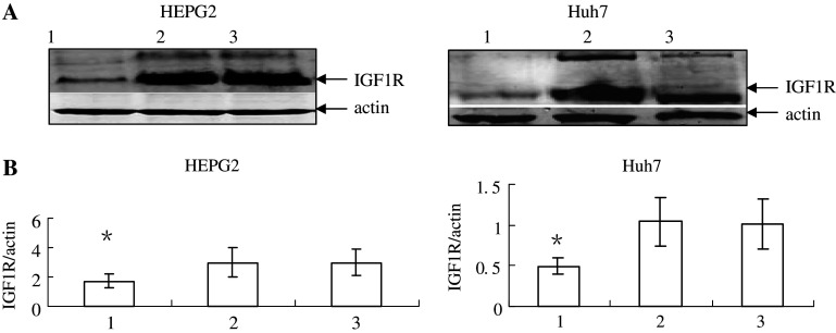
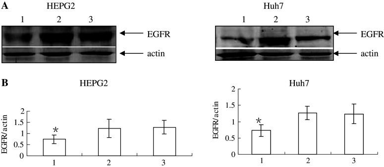
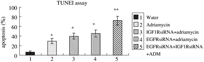
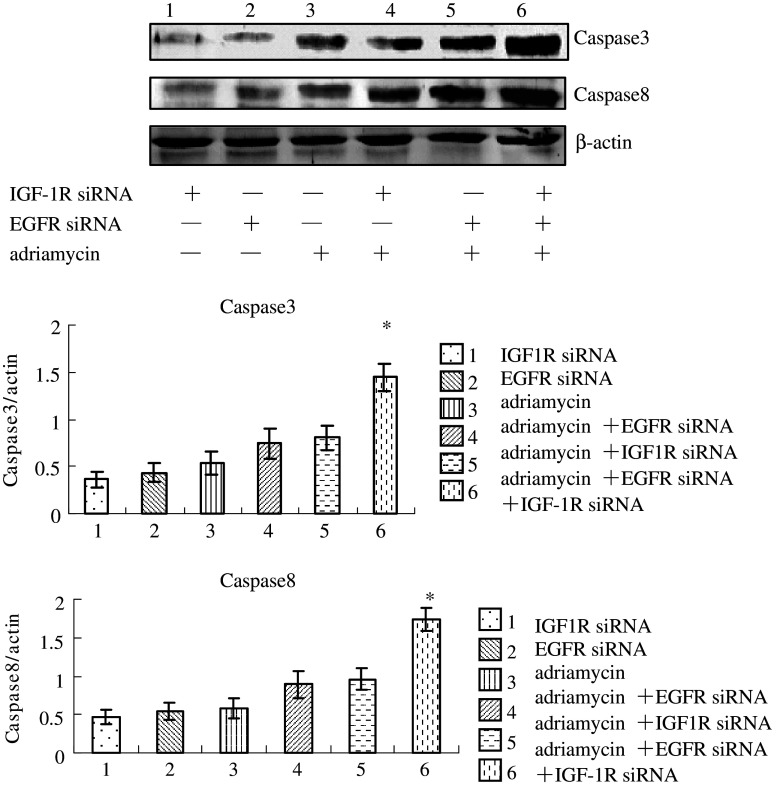
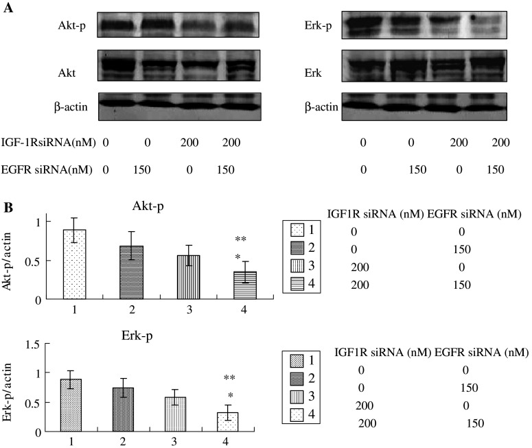
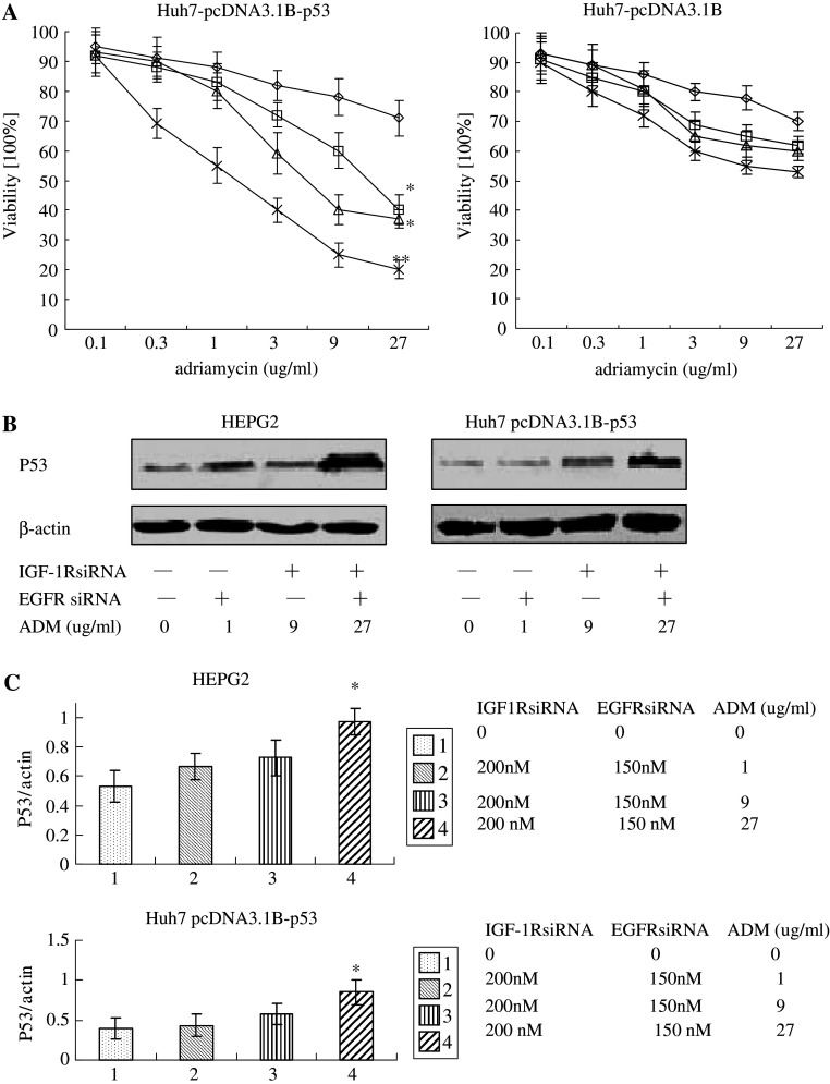
Methods: HEPG2, Huh7 cell lines were transfected IGF1R siRNAs and EGFR siRNAs and IGF1R or EGFR mRNA level was determined by RT-PCR and Western-blot analysis. We investigated the effects of the adriamycin-induced apoptosis of these cells by TUNEL assay. Also we analyze caspase3, 8 and the phosphorylation levels of Akt and Erk by Western-blot. The p53 effect of adriamycin-induced cell death by inhibitors of EGFR/IGF1R is investigated by cell growth curves.

Results: Transfection of an IGF1R and EGFR siRNAs resulted in substantial loss of IGF1R and EGFR mRNA of HEPG2, Huh7 cells relative to the control case. EGFR siRNA and IGF1R siRNA treatments increased the adriamycin-induced apoptosis of these cells. IGF1R siRNA and EGFR siRNA enhance a caspase-dependent cell death program. The phosphorylation levels of Akt and Erk were reduced by the combination of the two agents. The facilitation of adriamycin-induced cell death by inhibitors of EGFR/IGF1R is p53-independent.

Conclusions: The results indicate that the siRNA for IGF1R has a great potential for cancer therapy when combined with either a chemotherapeutic agent or siRNAs that targets EGFR.


Figures

Fig. 1
Fig. 1
HEPG2 and Huh7 cells were treated with siRNA duplex targeting IGF1R (50 nM), EGFR (150 nM), scramble or distilled water and then 48 h later, cell proteins were analyzed for IGF1R by Western-blot analysis as described under Materials and methods. 1 anti-IGF1R; 2 anti-scramble; 3 water; effects of siRNAs on IGF1R protein in HEPG2 and Huh7 cells. Results are means ± SD for three replicate determinations for each treatment group, and a significant (P < 0.05) decrease in IGF1R proteins levels was observed only in cells treated with siRNA for IGF1R. *P < 0.05 versus cells treated with water or scramble siRNA
Fig. 2
Fig. 2
HEPG2 and Huh7 cells were treated with siRNA duplex targeting IGF1R (50 nM), EGFR (150 nM), scramble or distilled water and then 48 h later, cell proteins were analyzed for EGFR proteins by Western-blot analysis as described under Materials and methods. 1 anti-EGFR; 2 anti-scramble; 3 water; effects of siRNAs on EGFR protein in HEPG2 and Huh7 cells. Results are means ± SD for three replicate determinations for each treatment group, and a significant (P < 0.05) decrease in EGFR proteins levels was observed only in cells treated with siRNA for EGFR. *P < 0.05 versus cells treated with water or scramble siRNA
Fig. 3
Fig. 3
IGF-1R inhibition synergistically enhances the proapoptotic effects of EGFR inhibition. HEPG2 cells were co-incubated with water, EGFR siRNA (150 nM), IGF-1R siRNA (50, 100, 200 nM), or their combination and exposed to adriamycin for 24 h. Note that IGF-1R siRNA alone does not modulate adriamycin-induced apoptosis at 100 nM (b), but still enhances the effect of EGFR siRNA. Viability was assessed by MTT assay (n = 3, SD). *P < 0.05 versus cells only treated with adriamycin. **P < 0.05 vs. cells treated with adriamycin after transfection with siRNA duplex targeting IGF-1R or EGFR alone. open diamond water after distilled water; open square IGF-1R siRNA; open triangle EGFR siRNA; multiplication symbol EGFR siRNA + IGF-1R siRNA
Fig. 4
Fig. 4
Programmed cell death in HEPG2 cells transfected with the siRNAs for IGF1R (200 nM) and EGFR (150 nM) for 48 h, and then treated with adriamycin(27 μg/ml) for 24 h detected by the TUNEL assay. Results are represented as a % of all the cells, considering this as 100%. It is possible to observe increased apoptosis in cells treated with the siRNAs for IGF1R or/and EGFR. Results are the mean of three independent experiments (each with three replicates). *P < 0.05 versus cells treated with water. **P < 0.05 versus cells treated with adriamycin
Fig. 5
Fig. 5
Co-inhibition of EGFR and IGF-1R enhances adriamycin-induced cell death in a caspase-dependent fashion. HEPG2 cells were incubated with water, EGFR siRNA (150 nM), IGF-1R siRNA (200 nM) or both and exposed to adriamycin (27 μg/ml) for 24 h. The expression of caspase 3, 8 was detected by Western-blot. *P < 0.05 versus cells treated with adriamycin
Fig. 6
Fig. 6
Effect of treatment on Erk and Akt kinases phosphorylation and protein levels. Western blot analysis showing phosphorylation (P) and protein levels of Akt and p44/p42 Erk kinases in cells treated for 48 h with IGF-1RsiRNA or/and EGFR siRNA. *P < 0.05 versus cells treated with water. **P < 0.05 versus cells transfected with siRNA duplex targeting IGF-1R or EGFR alone
Fig. 7
Fig. 7
The facilitation of adriamycin-induced cell death by inhibitors of EGFR/IGF-1R is p53-independent. a Huh7 pcDNA3.1B cells (possessing mutant p53), Huh7 pcDNA3.1B-p53 cells (possessing wild-type p53) were incubated with EGFR siRNA (150 nM), IGF-1RsiRNA (200 nM) or both and exposed to adriamycin for 24 h. Viability was assessed by MTT (n = 3, SD). b Western-blotting analysis for expression of P53 in HEPG2 and Huh7 pcDNA3.1B-p53 cells lines (both possessing wild-type P53). Two cell lines incubated with EGFRsiRNA (150 nM), IGF-1RsiRNA (200 nM) or both and exposed to adriamycin for 24 h. Actin served as a control. Treatment, harvest and analysis were repeated three times. *P < 0.05 versus cells only treated with adriamycin. **P < 0.05 versus cells treated with adriamycin after transfection with siRNA duplex targeting IGF-1R or EGFR alone

References

    1. Aden DP, Fogel A, Plotkin S, Damjanov I, Knowles BB (1979) Controlled synthesis of HBsAg in a differentiated human liver carcinoma-derived cell line. Nature 282:615–616 - PubMed
    1. Albanell J, Baselga J (1996) Unraveling resistance to trastuzuMAb (Herceptin): insulin-like growth factor-I receptor, a new suspect. J Natl Cancer Inst 93:1830–1832 - PubMed
    1. Anderson NG, Ahmad T, Chan K, Dobson R, Bundred NJ (2001) ZD1839 (Iressa), a novel epidermal growth factor receptor (EGFR) tyrosine kinase inhibitor, potently inhibits the growth of EGFRpositive cancer cell lines with or without erbB2 overexpression. Int J Cancer 94:774–782 - PubMed
    1. Aplen NJ, Parrish S, Imani F, Fire A, Morgan RA (2001) Specific inhibition of gene expression by small double-stranded RNAs in invertebrate and vertebrate systems. Proc Natl Acad Sci USA 98:9742–9747 - PMC - PubMed
    1. Arteaga C (2002) Epidermal growth factor receptor dependence in human tumors: more than just expression? Oncologist 7:31–39 - PubMed

Publication types

MeSH terms