

Digital dermoscopic monitoring of atypical nevi in patients at risk for melanoma
- PMID: 17903152
- PMCID: PMC2292405
- DOI: 10.1111/j.1524-4725.2007.33254.x
Digital dermoscopic monitoring of atypical nevi in patients at risk for melanoma
Abstract
Background: Atypical nevi are a common risk factor for melanoma.
Objectives: The objective was to determine the utility of monitoring dermoscopic photographs of atypical nevi in a high-risk population.
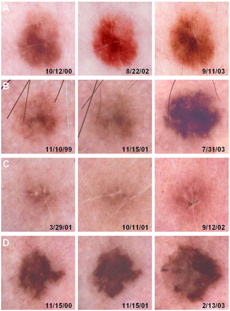
Methods: Over a 4.5-year period, digital dermoscopic photographs were taken of clinically atypical nevi at initial and follow-up visits, such that side-by-side comparisons could be made.
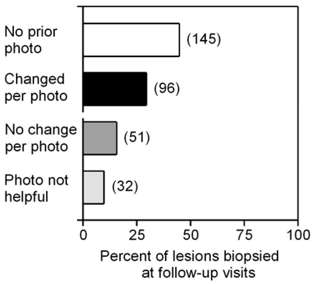
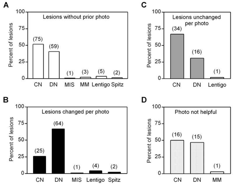
Results: A total of 5,945 lesions were monitored in 297 patients over 3 to 52 months (median, 22 months), and 324 lesions were biopsied. Photographic (dermoscopic) changes were noted in 96 of 5,945 (1.6%) lesions, which included 64 dysplastic nevi (67%), 25 common nevi (26%), and 1 melanoma (1.0%). Of 6 melanomas biopsied during the follow-up period, only 1 was detected by dermoscopic photographic change at follow-up.
Conclusions: Most clinically atypical melanocytic nevi are stable over time, and lesions exhibiting dermoscopic changes are most likely to be dysplastic nevi. Although dermoscopy is a useful tool for clinical examination, the sensitivity of dermoscopic monitoring is limited by melanomas that may arise in normal skin or in clinically benign nevi that were not initially photographed.
Conflict of interest statement
Stanley R. Fuller, BS, Glen M. Bowen, MD, Ben Tanner, BS, Scott R. Florell, MD, and Douglas Grossman, MD, PhD have no significant financial conflicts of interest.
Figures
Similar articles
-
Clinical and Histopathologic Characteristics of Melanocytic Lesions on the Volar Skin Without Typical Dermoscopic Patterns.JAMA Dermatol. 2019 May 1;155(5):578-584. doi: 10.1001/jamadermatol.2018.5926. JAMA Dermatol. 2019. PMID: 30865233 Free PMC article.
-
Dermoscopy for the pediatric dermatologist part III: dermoscopy of melanocytic lesions.Pediatr Dermatol. 2013 May-Jun;30(3):281-93. doi: 10.1111/pde.12041. Epub 2012 Dec 18. Pediatr Dermatol. 2013. PMID: 23252411 Review.
-
[Thermographic examination of cutaneous melanocytic nevi].Ann Acad Med Stetin. 2009;55(1):31-8; discussion 38. Ann Acad Med Stetin. 2009. PMID: 20349589 Polish.
-
The diagnostic value and histologic correlate of distinct patterns of shiny white streaks for the diagnosis of melanoma: A retrospective, case-control study.J Am Acad Dermatol. 2018 May;78(5):913-919. doi: 10.1016/j.jaad.2017.11.021. Epub 2017 Nov 11. J Am Acad Dermatol. 2018. PMID: 29138058 Free PMC article.
-
Dermoscopic features of common nevi: a review.G Ital Dermatol Venereol. 2012 Apr;147(2):141-8. G Ital Dermatol Venereol. 2012. PMID: 22481577 Review.
Cited by
-
Outpatient Follow-up and Secondary Prevention for Melanoma Patients.Cancers (Basel). 2010 Jun 7;2(2):1178-97. doi: 10.3390/cancers2021178. Cancers (Basel). 2010. PMID: 24281112 Free PMC article.
-
Clinical performance of the Nevisense system in cutaneous melanoma detection: an international, multicentre, prospective and blinded clinical trial on efficacy and safety.Br J Dermatol. 2014 Nov;171(5):1099-107. doi: 10.1111/bjd.13121. Epub 2014 Oct 19. Br J Dermatol. 2014. PMID: 24841846 Free PMC article.
-
The dysplastic nevus: from historical perspective to management in the modern era: part I. Historical, histologic, and clinical aspects.J Am Acad Dermatol. 2012 Jul;67(1):1.e1-16; quiz 17-8. doi: 10.1016/j.jaad.2012.02.047. J Am Acad Dermatol. 2012. PMID: 22703915 Free PMC article.
-
Benefits of total body photography and digital dermatoscopy ("two-step method of digital follow-up") in the early diagnosis of melanoma in patients at high risk for melanoma.J Am Acad Dermatol. 2012 Jul;67(1):e17-27. doi: 10.1016/j.jaad.2011.04.008. Epub 2011 Jun 16. J Am Acad Dermatol. 2012. PMID: 21683472 Free PMC article.
-
Definition of an automated Content-Based Image Retrieval (CBIR) system for the comparison of dermoscopic images of pigmented skin lesions.Biomed Eng Online. 2009 Aug 16;8:18. doi: 10.1186/1475-925X-8-18. Biomed Eng Online. 2009. PMID: 19682395 Free PMC article.
References
-
- Brochez L, Verhaeghe E, Bleyen L, Naeyaert JM. Diagnostic ability of general practitioners and dermatologists in discriminating pigmented skin lesions. J Am Acad Dermatol. 2001;44:979–86. - PubMed
-
- Binder M, Schwarz M, Winkler A, Steiner A, Kaider A, Wolff K, Pehamberger H. Epiluminescence microscopy. A useful tool for the diagnosis of pigmented skin lesions for formally trained dermatologists. Arch Dermatol. 1995;131:286–91. - PubMed
-
- Carli P, de Giorgi V, Chiarugi A, Nardini P, Weinstock MA, Crocetti E, Stante M, Giannotti B. Addition of dermoscopy to conventional naked-eye examination in melanoma screening: a randomized study. J Am Acad Dermatol. 2004;50:683–9. - PubMed
-
- Dolianitis C, Kelly J, Wolfe R, Simpson P. Comparative performance of 4 dermoscopic algorithms by nonexperts for the diagnosis of melanocytic lesions. Arch Dermatol. 2005;141:1008–14. - PubMed
-
- Rigel DS, Friedman RJ, Kopf AW, Polsky D. ABCDE--an evolving concept in the early detection of melanoma. Arch Dermatol. 2005;141:1032–4. - PubMed
Publication types
MeSH terms
Grants and funding
LinkOut - more resources
Full Text Sources
Medical
