

Immunohistological assessment of the synovial tissue in small joints in rheumatoid arthritis: validation of a minimally invasive ultrasound-guided synovial biopsy procedure
- PMID: 17903238
- PMCID: PMC2212566
- DOI: 10.1186/ar2302
Immunohistological assessment of the synovial tissue in small joints in rheumatoid arthritis: validation of a minimally invasive ultrasound-guided synovial biopsy procedure
Abstract
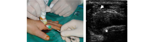
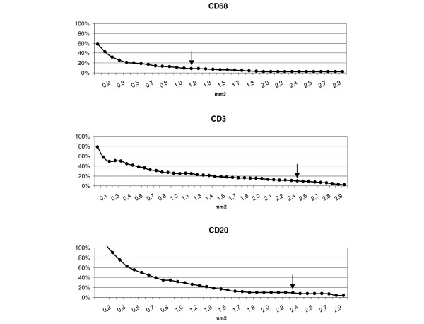
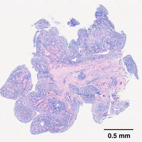
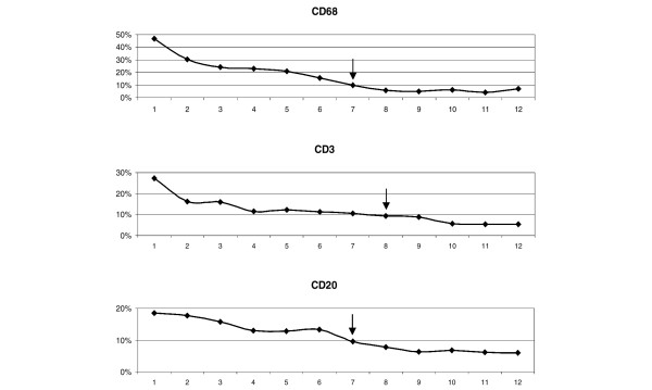
The aim of the present study was to perform an immunohistological assessment of the synovial tissue from involved small joints in rheumatoid arthritis (RA) and to explore the reliability of a mini-invasive ultrasound (US)-guided technique of small joint synovial biopsy for the histopathological assessment. Synovial tissue collected during arthrotomic surgery of small joints in nine patients served as the gold standard for the validation of the histological assessment. Small hand-joint synovial biopsies from an additional nine patients with erosive RA were obtained by a mini-invasive US-guided procedure, performed percutaneously by the portal and rigid forceps technique. Using digital image analysis, the area fractions of synovial macrophages (CD68 cells), T cells (CD3 cells) and B cells (CD20 cells) were measured in all high-power fields of every sample at different cutting levels. The representative sample was defined as the minimal number of high-power fields whose mean area fraction would reflect the overall mean area fraction within a percentage mean difference of 10%. For each patient, a range of three to five large samples for surgical biopsies and a range of 8-12 samples for US-guided biopsies were collected and analysed. In arthrotomic samples, the analysis of a randomly selected tissue area of 2.5 mm2 was representative of the overall value for CD68, CD3 and CD20 cells. US-guided samples allowed histological evaluation in 100% of cases, with a mean valid area of 18.56 mm2 (range 7.29-38.28 mm2). The analysis of a cumulative area of 2.5 mm2 from eight randomly selected sections (from different samples or from different cutting levels) allowed to reduce the percentage mean difference to less than 10% for CD68, CD3 and CD20 cells. In conclusion, US-guided synovial biopsy represents a reliable tool for the assessment of the histopathological features of RA patients with a mini-invasive approach.
Figures
Similar articles
-
Ultrasound-guided synovial biopsy: a safe, well-tolerated and reliable technique for obtaining high-quality synovial tissue from both large and small joints in early arthritis patients.Ann Rheum Dis. 2015 Mar;74(3):611-7. doi: 10.1136/annrheumdis-2013-204603. Epub 2013 Dec 13. Ann Rheum Dis. 2015. PMID: 24336336
-
A Multicenter Retrospective Analysis Evaluating Performance of Synovial Biopsy Techniques in Patients With Inflammatory Arthritis: Arthroscopic Versus Ultrasound-Guided Versus Blind Needle Biopsy.Arthritis Rheumatol. 2018 May;70(5):702-710. doi: 10.1002/art.40433. Epub 2018 Apr 2. Arthritis Rheumatol. 2018. PMID: 29409140
-
Use of ultrasound-guided small joint biopsy to evaluate the histopathologic response to rheumatoid arthritis therapy: recommendations for application to clinical trials.Arthritis Rheumatol. 2015 Oct;67(10):2601-10. doi: 10.1002/art.39235. Arthritis Rheumatol. 2015. PMID: 26097225
-
Evaluation of Minimally Invasive, Ultrasound-guided Synovial Biopsy Techniques by the OMERACT Filter--Determining Validation Requirements.J Rheumatol. 2016 Jan;43(1):208-13. doi: 10.3899/jrheum.141199. Epub 2015 Jun 1. J Rheumatol. 2016. PMID: 26034155 Review.
-
Best practices for ultrasound-guided synovial biopsy in the United States.Best Pract Res Clin Rheumatol. 2023 Mar;37(1):101834. doi: 10.1016/j.berh.2023.101834. Epub 2023 May 30. Best Pract Res Clin Rheumatol. 2023. PMID: 37263809 Review.
Cited by
-
Utility of synovial biopsy.Arthritis Res Ther. 2009;11(6):256. doi: 10.1186/ar2847. Epub 2009 Nov 23. Arthritis Res Ther. 2009. PMID: 19951395 Free PMC article. Review.
-
Expression of chemokines CXCL4 and CXCL7 by synovial macrophages defines an early stage of rheumatoid arthritis.Ann Rheum Dis. 2016 Apr;75(4):763-71. doi: 10.1136/annrheumdis-2014-206921. Epub 2015 Apr 9. Ann Rheum Dis. 2016. PMID: 25858640 Free PMC article.
-
Ultrasound-guided synovial biopsy improves diagnosis of septic arthritis in acute arthritis without enough analyzable synovial fluid: a retrospective analysis of 176 arthritis from a French rheumatology department.Clin Rheumatol. 2018 Aug;37(8):2241-2249. doi: 10.1007/s10067-018-4160-9. Epub 2018 Jun 14. Clin Rheumatol. 2018. PMID: 29948353
-
Ultrasound-Guided Synovial Biopsy: A Review.Front Med (Lausanne). 2021 Apr 22;8:632224. doi: 10.3389/fmed.2021.632224. eCollection 2021. Front Med (Lausanne). 2021. PMID: 33968950 Free PMC article. Review.
-
Inflammatory lesions in the bone marrow of rheumatoid arthritis patients: a morphological perspective.Arthritis Res Ther. 2012 Dec 27;14(6):229. doi: 10.1186/ar4115. Arthritis Res Ther. 2012. PMID: 23270711 Free PMC article. Review.
