Review
. 2007 Sep 18;8 Suppl 3(Suppl 3):S4.
doi: 10.1186/1471-2202-8-S3-S4.

Factors that regulate embryonic gustatory development

Affiliations
Review

Factors that regulate embryonic gustatory development

Robin F Krimm. BMC Neurosci. .

Abstract

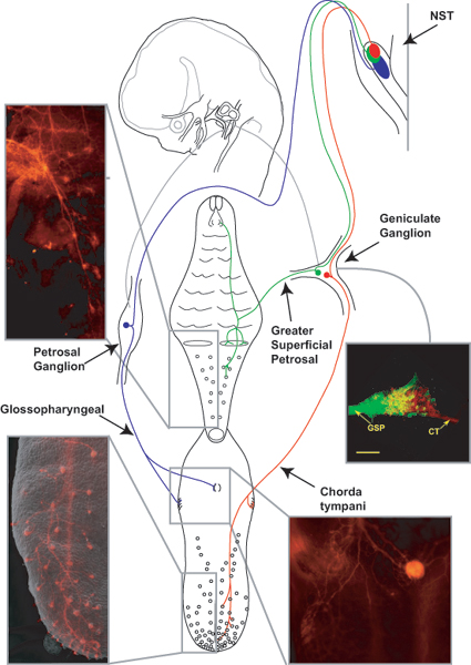
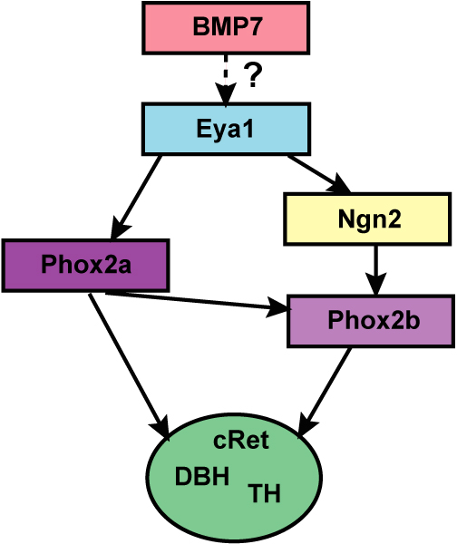
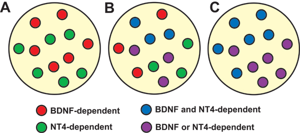
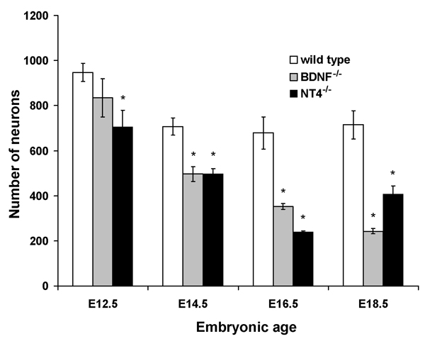
Numerous molecular factors orchestrate the development of the peripheral taste system. The unique anatomy/function of the taste system makes this system ideal for understanding the mechanisms by which these factors function; yet the taste system is underutilized for this role. This review focuses on some of the many factors that are known to regulate gustatory development, and discusses a few topics where more work is needed. Some attention is given to factors that regulate epibranchial placode formation, since gustatory neurons are thought to be primarily derived from this region. Epibranchial placodes appear to arise from a pan-placodal region and a number of regulatory factors control the differentiation of individual placodes. Gustatory neuron differentiation is regulated by a series of transcription factors and perhaps bone morphongenic proteins (BMP). As neurons differentiate, they also proliferate such that their numbers exceed those in the adult, and this is followed by developmental death. Some of these cell-cycling events are regulated by neurotrophins. After gustatory neurons become post-mitotic, axon outgrowth occurs. Axons are guided by multiple chemoattractive and chemorepulsive factors, including semaphorins, to the tongue epithelium. Brain derived neurotrophic factor (BDNF), functions as a targeting factor in the final stages of axon guidance and is required for gustatory axons to find and innervate taste epithelium. Numerous factors are involved in the development of gustatory papillae including Sox-2, Sonic hedge hog and Wnt-beta-catenin signaling. It is likely that just as many factors regulate taste bud differentiation; however, these factors have not yet been identified. Studies examining the molecular factors that regulate terminal field formation in the nucleus of the solitary tract are also lacking. However, it is possible that some of the factors that regulate geniculate ganglion development, outgrowth, guidance and targeting of peripheral axons may have the same functions in the gustatory CNS.


Figures

Figure 1
Figure 1
An overview of the basic neuroanatomy of the gustatory system. A cartoon of geniculate neurons innervating the tongue (red) and the palate (green) and petrosal neurons innervating the tongue (blue) are shown innervating peripheral taste bud containing regions and the rostral nucleus of the solitary tract (NST). On the tongue, taste buds are located in fungiform papillae, foliate papillae, and circumvallate papillae (CV). The palate has taste buds on the nasoincisor papilla/ducts (NID) and on the soft palate (circles). Photomicrographs of innervation patterns in the tongue and in the palate at E16.5 are shown next to the appropriate regions. An overlay image of two geniculate ganglia (E14.5) is also shown; one ganglia following DiI-label to the palate was pseudo-colored green, the other following DiI-labeling of the tongue remains red. These two ganglia images were anatomically aligned and superimposed using Adobe Photoshop.
Figure 2
Figure 2
During development, the early differentiation of the geniculate and nodose ganglia is regulated by a series of transcription factors and signals from the pharyngeal pouch endoderm. The Six and Eya families of transcription factors are important for the development of multiple placodes, including the epibranchial placodes, from a single, common placode. In this pan-placodal area, Pax2 expression demarcates a region that forms the epibranchial and otic placodes. Signals from the pharyngeal pouch endoderm, like members of the bone morphogenic protein family (BMP), are required to induce epibranchial placode formation. Also, neurogenin 2 (Ngn2) and Phox2a signaling are important for neuronal differentiation within the placodes. Both are dependent on Eya1, but independent of one other. Phox2b is dependent on both Phox2a and Ngn2. Phox2 genes may be important for general neuronal differentiation as well as for differentiation of neuron subtypes. We propose that an unidentified factor(s) regulates the differentiation of gustatory phenotype and subtypes within these ganglia.
Figure 3
Figure 3
Bdnf-/- andNtf5-/- mice lose 50% of geniculate/petrosal and nodose neurons. Mice lacking both BDNF and NT4/5 lose almost all of the neurons in these ganglia. At least two different scenarios could explain these findings. Two separate subpopulations could exist. One that is BDNF-dependent and one is that is NT4/5-dependent (A). In this case, BDNF dependent neurons would be lost in Bdnf-/- mice and NT4/5-dependent neurons are lost in Ntf5-/- mice. Alternatively, one subpopulation of geniculate/petrosal/nodose neurons may be dependent on both BDNF and NT4/5 (C) such that removal of either neurotrophin would result in death. These two possibilities are not mutually exclusive. That is, all four types of neuron dependencies could be present in the same ganglion (B).
Figure 4
Figure 4
Neurons are lost throughout embryonic development in Bdnf-/- and Ntf5-/- mice. At E12.5, the geniculate ganglion is still fused with the vestibular-cochlear ganglion, which explains the greater number of neurons in wild type mice at this age. Neurons are lost by E12.5 in Ntf5-/- mice, indicating that NT4/5-dependency begins earlier in embryonic development than does BDNF-dependency. In Bdnf-/- mice, neurons are first lost between E12.5 and E14.5. Losses continue to be greater in these animals, compared to wild type, throughout the remainder of embryonic development.
Figure 5
Figure 5
By E14.5, chorda tympani fibers have innervated fungiform papillae. Chorda tympani fibers have a stereotypical branching pattern in the tongue (A). A higher magnification view of the area outlined in (A) illustrates that each fiber bundle ends in a distinctive bulb shape known as a neural bud (B, arrows). Neural buds form when gustatory fibers penetrate the epithelium at the surface of a fungiform papilla (C). All papillae appear to be innervated when the image from a portion of a E16.5 tongue following DiI-labeling (D) and the SEM image from the same tongue region (F) are overlaid (E). However, there are some neural buds in locations where no fungiform papillae are present (arrows in E and F).
Figure 6
Figure 6
DiI-labeling reveals that normal innervation patterns (A, D) are disrupted by overexpression of BDNF (B, E) and loss of BDNF (C, F). By the first day of targeting (E14.5), wild type mice have very stereotyped innervation patterns, and each fiber bundle branch ends in a neural bud (A). Overexpression of BDNF throughout the epithelium increases branching and disrupts normal targeting (B, E). Very few neural buds are initially formed (B) and by E18.5 (E), few fungiform papillae are innervated. Innervation patterns are even more disrupted in Bdnf-/- mice. Axonal branching is extensive near the epithelial surface and neural buds fail to form (C). By E18.5, there is a loss of innervation throughout most of the tongue; however, a few neural buds were present, indicating that some fungiform papillae were innervated. Scale bar in A = 250 μm, applies to A-C; Scale bar in D = 500 μm, applies to D-F.
Figure 7
Figure 7
Small groups of taste cells are labeled with anti-troma I (anti-keratin 8) as early as E16.5 (A). These clusters increase in size and number by E18.5 (B) and by birth (C). Scale bar in F = 10 μm, applies to A-C.

References

    1. Barlow LA. Gustatory System Development. In: Finger TE, Silver WL, Restrepo D, editor. The Neurobiology of Taste and Smell. New York: Wiley-Liss; 2000. pp. 395–422.
    1. Northcutt RG, Barlow LA, Braun CB, Catania KC. Distribution and innervation of taste buds in the axolotl. Brain, behavior and evolution. 2000;56:123–145. doi: 10.1159/000047200. - DOI - PubMed
    1. Barlow LA. Toward a unified model of vertebrate taste bud development. J Comp Neurol. 2003;457:107–110. doi: 10.1002/cne.10549. - DOI - PubMed
    1. Hill DL, Mistretta CM. Developmental neurobiology of salt taste sensation. Trends Neurosci. 1990;13:188–195. doi: 10.1016/0166-2236(90)90046-D. - DOI - PubMed
    1. Mistretta CM, Hill DL. Development of the taste system: basic neurobiology. In: RL D, editor. Handbook of Olfaction and Gustation. New York: Marcel Dekker; 1995. pp. 635–668.

Publication types