. 2007 Nov 1;311(1):124-35.
doi: 10.1016/j.ydbio.2007.08.023. Epub 2007 Aug 17.

Mouse R-spondin2 is required for apical ectodermal ridge maintenance in the hindlimb

Affiliations

Mouse R-spondin2 is required for apical ectodermal ridge maintenance in the hindlimb

Ju-Suk Nam et al. Dev Biol. .

Abstract

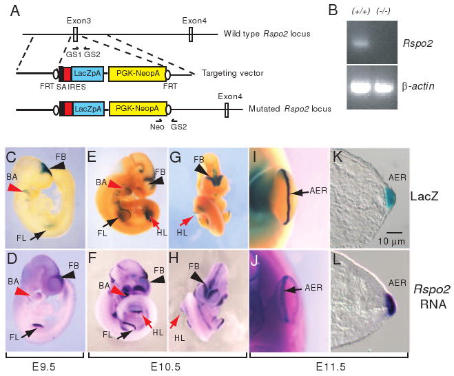
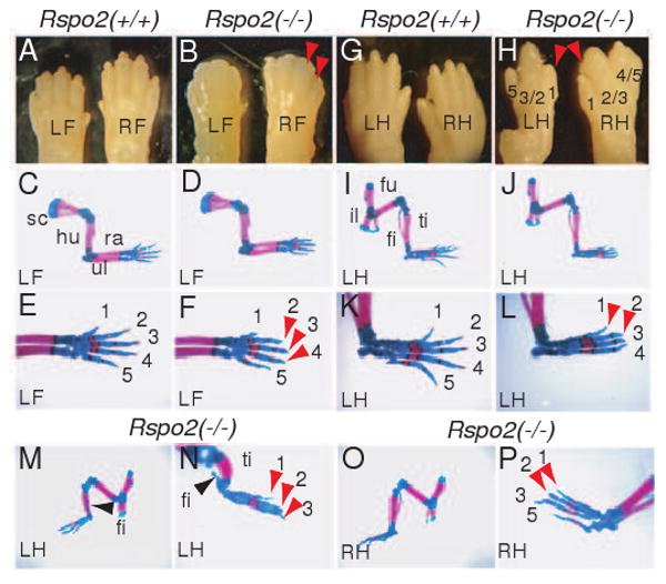
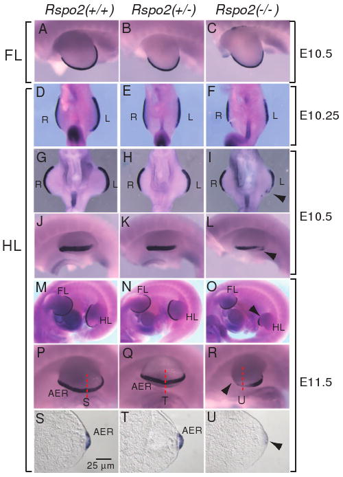
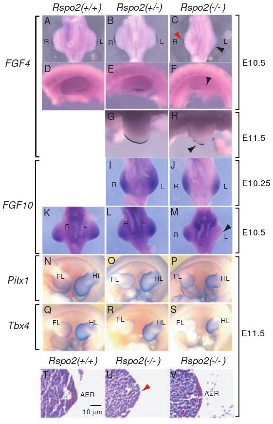
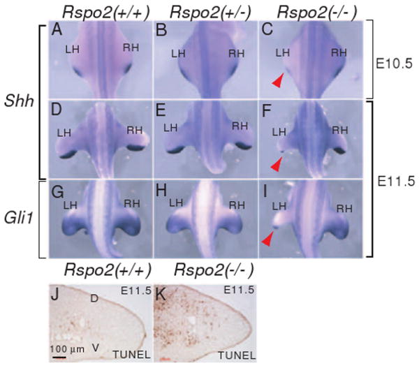
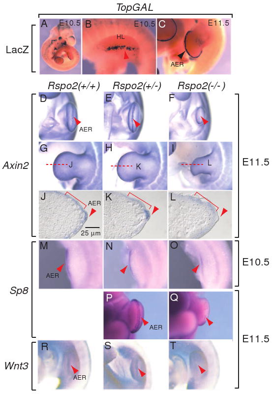
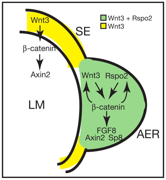
The R-spondin (Rspo) family of proteins consists of secreted cysteine-rich proteins that can activate beta-catenin signaling via the Frizzled/LRP5/6 receptor complex. Here, we report that targeted inactivation of the mouse Rspo2 gene causes developmental limb defects, especially in the hindlimb. Although the initiation of the expression of apical ectodermal ridge (AER)-specific genes, including fibroblast growth factor 8 (FGF8) and FGF4 occurred normally, the maintenance of these marker expressions was significantly defective in the hindlimb of Rspo2(-/-) mice. Consistent with the ligand role of R-spondins in the Wnt/beta-catenin signaling pathway, expression of Axin2 and Sp8, targets for beta-catenin signaling, within AER was greatly reduced in Rspo2(-/-) embryos. Furthermore, sonic hedgehog (Shh) signaling within the hindlimbs of Rspo2(-/-) mice was also significantly decreased. Rspo2 is expressed in the AER of all limb buds, however the stunted phenotype is significantly more severe in the hindlimbs than the forelimbs and strongly biased to the left side. Our findings strongly suggest that Rspo2 expression in the AER is required for AER maintenance likely by regulating Wnt/beta-catenin signaling.


Figures

Figure 1
Figure 1
Generation of Rspo2 gene-targeted mice. (A) An IRES-LacZ-PGKneor cassette flanked by a splicing acceptor (SA) was replaced with exon 3 of the mouse Rspo2 gene. Arrows indicate the positions of GS1, GS2, and Neo primers for genotyping. Black ovals indicate Flp-recombinase target sites (FRT). (B) Rspo2 RNA expression was not detected in Rspo2(-/-) mice. PCR primers specific for exon 2 and exon 5 were used to detect Rspo2 mRNA spanning the predicted coding region. Total RNA isolated from brain tissue of P1 (postnatal day 1) mice was used to synthesize cDNA templates. β-actin RNA expression is shown as control. LacZ expression (C, E, G, I and K) in Rspo2(+/-) mouse embryo and Rspo2 RNA expression in wild type mouse embryo (D, F, H, J and L) at embryonic day 9.5 (E9.5, C and D), E10.5 (E-H) and E11.5 (I-L). Abbreviations: FB, forebrain (black arrowheads); FL, forelimb bud (black arrows); HL, hindlimb bud (red arrows); BA, branchial arch (red arrowheads); AER, apical ectodermal ridge (black arrows in I and J).
Figure 2
Figure 2
Limb skeletal defects in Rspo2(-/-) mice. (A and B) Gross morphology of the distal forelimbs of Rspo2(+/+) and Rspo2(-/-) mice at P1. The right (RF) and left (LF) feet are indicated. Red arrowheads indicate the absence of claws in Rspo2(-/-) mouse. (C-F) Whole mount forelimb skeleton of Rspo2(+/+) and Rspo2(-/-) mouse fetuses at E18.5. Skeleton of the left forelimbs are presented. A magnified view of the foot skeleton shows the absence of ossification of the most distal phalangeal bones (indicated by red arrowheads in F) in Rspo2(-/-) mouse. (G and H) Gross morphology of the distal hindlimbs of Rspo2(+/+) and Rspo2(-/-) mice at P1. The right (RH) and left (LH) feet are indicated. Red arrowheads indicate the missing claws. Fusions between digits 2 and 3, and digits 4 and 5 are shown in Rspo2(-/-) mouse. (I-P) Whole mount hindlimb skeleton of Rspo2(+/+) and Rspo2(-/-) mice at E18.5. The skeleton of the left hindlimbs (LH) of Rspo2(-/-) showed mild defects (J and L). Note the absence of digit 5 and lack of ossification of the most distal phalangeal bones were detected. Hindlimb skeleton of Rspo2(-/-) mouse with more severe defects (M-P). The left hindlimb of Rspo2(-/-) mice (M and N) showed more severe defects than the right hindlimb of the same mice (O and P). The left hindlimb was photographed from the anterior side to better visualize the defect in the fibula (black arrowheads). Red arrowheads in N and P indicate the absence of ossification of the distal phalangeal bones. Abbreviations: fi, fibula; fu, femur; hu, humerus; il, ilium; ra, radius; sc, scapula; ti, tibia; ul, ulna.
Figure 3
Figure 3
FGF8 expression in the AER of the developing limbs of Rspo2(-/-) mouse embryos. FGF8 expression was analyzed by whole mount in situ hybridization in the forelimbs (A-C) and hindlimbs (D-R) of Rspo2(+/+), Rspo2(+/-), and Rspo2(-/-) embryos at E10.25 (D-F), E10.5 (A-C and G-L) and E11.5 (M-R). The left side (A-C and J-R) and ventral side (D-I) views are presented, respectively. The right limbs (R), left limbs (L), forelimbs (FL) and hindlimbs (HL) of mouse embryos are indicated. Black arrowheads indicate the reduced FGF8 expression. The red dotted lines indicate the sectioning planes for the sections presented in S-U. (S-U) Transverse sections of the hindlimbs showed reduced expression of FGF8 expression within the AER region of Rspo2(-/-) embryos at E11.5.
Figure 4
Figure 4
Gene expression in the developing limbs of Rspo2(-/-) mouse embryos. (A-H) FGF4 expression was analyzed by whole mount in situ hybridization in Rspo2(+/+), Rspo2(+/-), and Rspo2(-/-) embryos at E10.5 (A-F) and E11.5 (G-H). (I-M) FGF10 RNA expression was analyzed in Rspo2(+/+), Rspo2(+/-), and Rspo2(-/-) mouse embryos at E10.25 (I-J) and E10.5 (K-M), respectively. The ventral side (A-C and I-M) and left side views (D-H) of the hindlimbs are presented, respectively. The right (R) and left (L) limbs are indicated. Black and red arrowheads indicate the reduced FGF4 (C and H) and FGF10 (M) expression in the hindlimbs of Rspo2(-/-) mouse embryos. (N-S) RNA expression of the hindlimb-specific markers, Pitx1 (N-P) and Tbx4 (Q-S), in the left hindlimbs of E11.5 mouse embryos. The left side views are presented. The forelimbs (FL) and hindlimbs (HL) of mouse embryos are indicated. (T-V) Transverse paraffin sections of the hindlimbs stained with hematoxylin and eosin. The AER is outlined with a red dotted line. Red arrowhead in panel U indicates the absence of a discernable AER structure.
Figure 5
Figure 5
Disruption of Shh signaling in the ZPA of Rspo2(-/-) mouse embryos. (A-F) Shh RNA expression was analyzed in Rspo2(+/+), Rspo2(+/-) and Rspo2(-/-) mouse embryos at E10.5 (A-C) and E11.5 (D-F). (G-I) RNA expression of Gli1, a downstream target gene for Shh signaling. The dorsal views of embryos at the hindlimb level are presented. The left (LH) and right (RH) hindlimbs are indicated. Red arrowheads (C, F and I) indicate the reduced expression of Shh and Gli1 RNA expression in Rspo2(-/-) embryos. (J and K) Cell apoptosis was detected by TUNEL staining in the left hindlimbs of Rspo2(+/+) and Rspo2(-/-) mouse embryos at E11.5. Transverse sections along the proximal-distal axis were analyzed and the presented sections were from the posterior portion of the limbs.
Figure 6
Figure 6
Inhibition of Wnt/β-catenin signaling in the AER of the hindlimbs of Rspo2(-/-) mouse embryos. (A-C) LacZ expression in the AER of the left hindlimbs of TopGAL transgenic mouse embryos at E10.5 (A and B) and E11.5 (C). The left hindlimds (HL) are presented in B and C. (D-L) RNA expression of Axin2, a β-catenin target gene, in the AER of the left hindlimbs of Rspo2(+/+), Rspo2(+/-), and Rspo2(-/-) mouse embryos at E11.5. The ventral side (D-F) and leftside (G-I) views of the hindlimbs are presented. The red dotted lines indicate the sectioning planes for the sections presented in J-L. (J-L) Transverse sections of the left hindlimbs of mouse Rspo2 mutant embryos stained with Axin2 riboprobe. Red brackets and arrowheads indicate Axin2 expression in mesenchymal cells and the AER, respectively. (M-Q) RNA expression of Sp8, another β-catenin downstream gene, in the left hindlimbs of Rspo2(+/+), Rspo2(+/-), and Rspo2(-/-) embryos at E10.5 (M-O) and E11.5 (P-Q). (R-T) Wnt3 RNA expression in the left hindlimbs of Rspo2(+/+), Rspo2(+/-), and Rspo2(-/-) embryos at E11.5. Red arrowheads indicate the expression of markers in the AER.
Figure 7
Figure 7
An integrated model of Rspo2-induced signaling in the AER of the mouse hindlimb. In the AER, Rspo2 and Wnt3 may act together or independently to activate strong β-catenin signaling, which leads to the activation of FGF8, Sp8 and Axin2 genes. In contrast, in the surface ectoderm (SE) outside of the AER where no Rspo2 expresses, Wnt3 may activate Axin2 in limb mesenchymal (LM) cells likely via β-catenin signaling. Although Wnt3 alone may be an enough signal for Axin2 expression, Rspo2 may be essential to regulate FGF8 and Sp8 expression. Solid arrows indicate the regulatory interactions that were proposed in this study and previously determined.

References

    1. Aoki M, Mieda M, Ikeda T, Hamada Y, Nakamura H, Okamoto H. R-spondin3 is required for mouse placental development. Dev Biol. 2007;301:218–226. - PubMed
    1. Barrow JR, Thomas KR, Boussadia-Zahui O, Moore R, Kemler R, Capecchi MR, McMahon AP. Ectodermal Wnt3/beta-catenin signaling is required for the establishment and maintenance of the apical ectodermal ridge. Genes Dev. 2003;17:394–409. - PMC - PubMed
    1. Bell SM, Schreiner CM, Waclaw RR, Campbell K, Potter SS, Scott WJ. Sp8 is crucial for limb outgrowth and neuropore closure. Proc Natl Acad Sci U S A. 2003;100:12195–200. - PMC - PubMed
    1. Bergmann C, Senderek J, Anhuf D, Thiel CT, Ekici AB, Poblete-Gutierrez P, van Steensel M, Seelow D, Nurnberg G, Schild HH, Nurnberg P, Reis A, Frank J, Zerres K. Mutations in the gene encoding the Wnt-signaling component R-spondin 4 (RSPO4) cause autosomal recessive anonychia. Am J Hum Genet. 2006;79:1105–9. - PMC - PubMed
    1. Blaydon DC, Ishii Y, O'Toole EA, Unsworth HC, Teh MT, Ruschendorf F, Sinclair C, Hopsu-Havu VK, Tidman N, Moss C, Watson R, de Berker D, Wajid M, Christiano AM, Kelsell DP. The gene encoding R-spondin 4 (RSPO4), a secreted protein implicated in Wnt signaling, is mutated in inherited anonychia. Nat Genet. 2006;38:1245–7. - PubMed

Publication types

LinkOut - more resources