. 2007 Sep 29;370(9593):1137-45.
doi: 10.1016/S0140-6736(07)61515-3.

H5N1 infection of the respiratory tract and beyond: a molecular pathology study

Affiliations

H5N1 infection of the respiratory tract and beyond: a molecular pathology study

Jiang Gu et al. Lancet. .

Abstract

Background: Human infection with avian influenza H5N1 is an emerging infectious disease characterised by respiratory symptoms and a high fatality rate. Previous studies have shown that the human infection with avian influenza H5N1 could also target organs apart from the lungs.

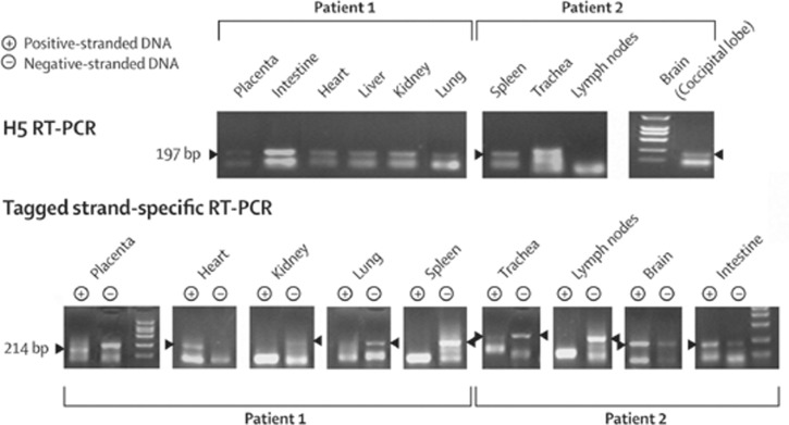
Methods: We studied post-mortem tissues of two adults (one man and one pregnant woman) infected with H5N1 influenza virus, and a fetus carried by the woman. In-situ hybridisation (with sense and antisense probes to haemagglutinin and nucleoprotein) and immunohistochemistry (with monoclonal antibodies to haemagglutinin and nucleoprotein) were done on selected tissues. Reverse-transcriptase (RT) PCR, real-time RT-PCR, strand-specific RT-PCR, and nucleic acid sequence-based amplification (NASBA) detection assays were also undertaken to detect viral RNA in organ tissue samples.

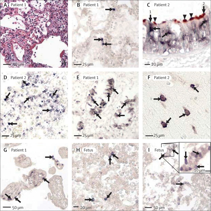
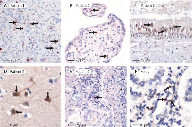
Findings: We detected viral genomic sequences and antigens in type II epithelial cells of the lungs, ciliated and non-ciliated epithelial cells of the trachea, T cells of the lymph node, neurons of the brain, and Hofbauer cells and cytotrophoblasts of the placenta. Viral genomic sequences (but no viral antigens) were detected in the intestinal mucosa. In the fetus, we found viral sequences and antigens in the lungs, circulating mononuclear cells, and macrophages of the liver. The presence of viral sequences in the organs and the fetus was also confirmed by RT-PCR, strand-specific RT-PCR, real-time RT-PCR, and NASBA.

Interpretation: In addition to the lungs, H5N1 influenza virus infects the trachea and disseminates to other organs including the brain. The virus could also be transmitted from mother to fetus across the placenta.


Figures

Figure 1
Figure 1
In-situ hybridisation locating gene sequences of H5N1 viral antigens (nucleoprotein and haemagglutinin) Signals seen with nitroblue tetrazolium/5-bromo-4-choloro-3-indolyl phosphate (purple-blue) and immunohistochemical signals with 3-amino-9-ethylcarbazole (double labelling, brown-red). No counterstaining done unless stated otherwise. (A) Lung tissue showing severe damage, hyaline membrane formation, and oedema (by haematoxylin and eosin staining), by contrast with limited number of cells positive for in-situ hybridisation in lung tissue of patient 1 (figure 1B). (B) Positive signals (with nucleoprotein sense probe) in nuclei of isolated pneumocytes (arrows). (C) Double labelling of in-situ hybridisation (with nucleoprotein antisense probe) and immunohistochemistry (with tubulin-β antibody; brown, arrowheads) showing dark-blue viral genomic sequences (arrows) in cytoplasm of tubulin-negative non-ciliated cell (arrow 1) and tubulin-positive ciliated cell (arrow 2) in trachea. No signal seen in nuclei (lightly counterstained with methyl-green). (D) Positive signals (with nucleoprotein sense probe) in several mononuclear cells in lymph node (arrows, lightly counterstained with methyl-green). (E) Positive signals (with nucleoprotein antisense probe) in mucosal epithelial cells of small intestine (arrows, lightly counterstained with methyl-green). (F) Positive signals (with nucleoprotein sense probe) in brain cells (arrows) from left parietal lobe (lightly counterstained with methyl-green), mainly located in cytoplasm and confirmed to be neurons from immunostaining for neurofilament or neuron-specific enolase (webfigures 1O and 1P). (G) Positive signals (with nucleoprotein antisense probe) in large mononuclear cells with morphological features of cytotrophoblasts in periphery of chorionic villus (arrows). Cells confirmed as cytotrophoblasts (webfigure 4D). No positive signals noted in any syncytiotrophoblasts. (H) Positive signals (with nucleoprotein sense probe) in fetal liver cells (arrows), confirmed as Kupffer cells (webfigure 1F). (I) Positive signals (with haemagglutinin sense probe) in bronchiolar epithelial cells in fetal lung tissue (arrows).
Figure 2
Figure 2
Immunohistochemical staining for H5N1 viral antigens (nucleoprotein, haemagglutinin) Positive signals seen with diaminobenzidine (brown; Zymed Laboratories, San Francisco, CA, USA) or 3-amino-9-ethylcarbazole (red-brown; Sigma, St Louis, MO, USA). Slides counterstained with haematoxylin. (A) Positive staining (with nucleoprotein antibody) in nuclei and cytoplasm of pneumocytes (arrows). (B) Positive signals (with nucleoprotein antibody) in nuclei of mononuclear cells (arrows) with morphological features of macrophages in core of chorionic villus. Immunostaining in adjacent sections indicate cells to be Hofbauer cells (webfigure 4). (C) Positive signals (with nucleoprotein antibody) in nuclei of epithelial cells of trachea (arrows). (D) Positive staining (with nucleoprotein antibody) in cytoplasm and nuclei of neurons (arrows) from hippocampus. (E) Positive staining (with haemagglutinin antibody) in mononuclear cell in lymph node (arrow). (F) Positive staining (with haemagglutin antibody) in cytoplasm of pneumocytes (arrows) in fetal lung tissue.
Figure 3
Figure 3
H5 RT-PCR and tagged strand-specific RT-PCR on selected organs No positive band in RT-PCR seen for lymph nodes in patient 2.

Comment in

References

    1. WHO Cumulative number of confirmed human cases of avian influenza A/(H5N1) reported to WHO. http://www.who.int/csr/disease/avian_influenza/country/cases_table_2007_... (accessed March 1, 2007).
    1. Ungchusak K, Auewarakul P, Dowell SF. Probable person-to-person transmission of avian influenza A (H5N1) N Engl J Med. 2005;352:333–340. - PubMed
    1. Normile D. Human transmission but no pandemic in Indonesia. Science. 2006;312:1855. - PubMed
    1. Horimoto T, Kawaoka Y. Pandemic threat posed by avian influenza A viruses. Clin Microbiol Rev. 2001;14:129–149. - PMC - PubMed
    1. Uiprasertkul MP, Puthavathana K, Sangsiriwut P. Influenza A H5N1 replication sites in humans. Emerg Infect Dis. 2005;11:1036–1041. - PMC - PubMed

Publication types

MeSH terms