. 2007;35(19):6611-24.
doi: 10.1093/nar/gkm612. Epub 2007 Sep 28.

Backbone-base inclination as a fundamental determinant of nucleic acid self- and cross-pairing

Affiliations

Backbone-base inclination as a fundamental determinant of nucleic acid self- and cross-pairing

Pradeep S Pallan et al. Nucleic Acids Res. 2007.

Abstract

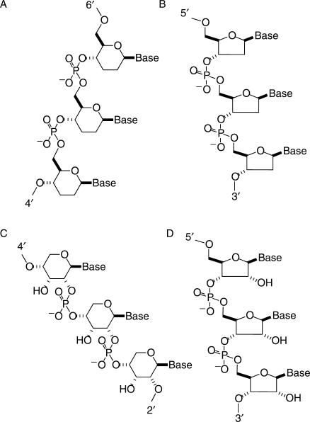
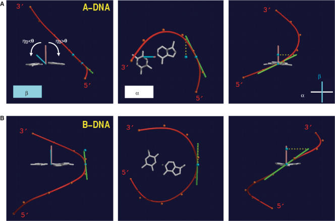
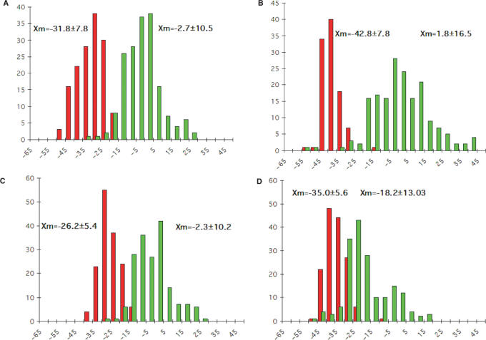
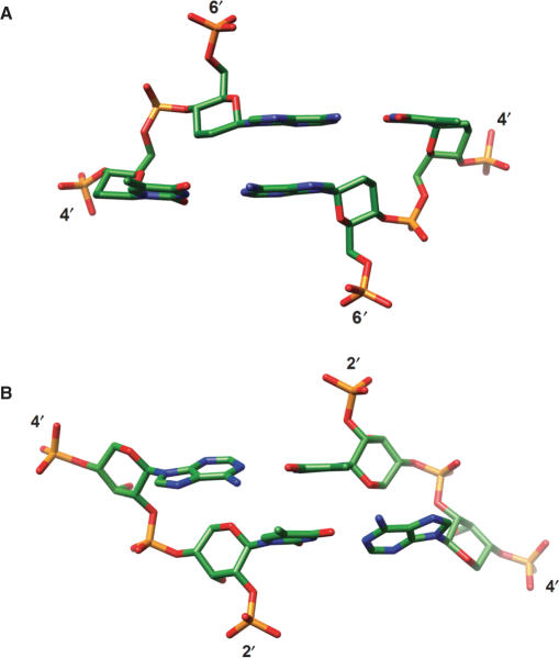
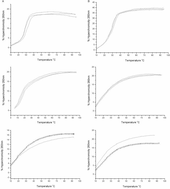
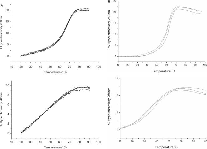
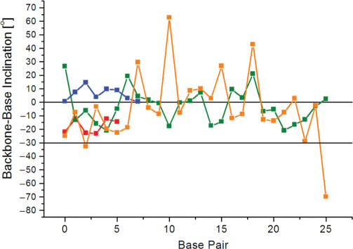
The crystal structure of the duplex formed by oligo(2',3'-dideoxy-beta-d-glucopyranosyl)nucleotides (homo-DNA) revealed strongly inclined backbone and base-pair axes [Egli,M., Pallan,P.S., Pattanayek,R., Wilds,C.J., Lubini,P., Minasov,G., Dobler,M., Leumann,C.J. and Eschenmoser,A. (2006) Crystal structure of homo-DNA and nature's choice of pentose over hexose in the genetic system. J. Am. Chem. Soc., 128, 10847-10856]. This inclination is easily perceived because homo-DNA exhibits only a modest helical twist. Conversely, the tight coiling of strands conceals that the backbone-base inclinations for A- (DNA and RNA) and B-form (DNA) duplexes differ considerably. We have defined a parameter eta(B) that corresponds to the local inclination between sugar-phosphate backbone and base plane in nucleic acid strands. Here, we show its biological significance as a predictive measure for the relative strand polarities (antiparallel, aps, or parallel, ps) in duplexes of DNA, RNA and artificial nucleic acid pairing systems. The potential of formation of ps duplexes between complementary 16-mers with eight A and U(T) residues each was investigated with DNA, RNA, 2'-O-methylated RNA, homo-DNA and p-RNA, the ribopyranosyl isomer of RNA. The thermodynamic stabilities of the corresponding aps duplexes were also measured. As shown previously, DNA is capable of forming both ps and aps duplexes. However, all other tested systems are unable to form stable ps duplexes with reverse Watson-Crick (rWC) base pairs. This observation illustrates the handicap encountered by nucleic acid systems with inclinations eta(B) that differ significantly from 0 degrees to form a ps rWC paired duplex. Accordingly, RNA with a backbone-base inclination of -30 degrees , pairs strictly in an aps fashion. On the other hand, the more or less perpendicular orientation of backbone and bases in DNA allows it to adopt a ps rWC paired duplex. In addition to providing a rationalization of relative strand polarity with nucleic acids, the backbone-base inclination parameter is also a determinant of cross-pairing. Thus, systems with strongly deviating eta(B) angles will not pair with each other. Nucleic acid pairing systems with significant backbone-base inclinations can also be expected to display different stabilities depending on which terminus carries unpaired nucleotides. The negative inclination of RNA is consistent with the higher stability of duplexes with 3'- compared to those with 5'-dangling ends.


Figures

Figure 1.
Figure 1.
Constitution and configuration of native and artificial nucleic acid pairing systems. (A) β-d-2′,3′-dideoxyglucopyranosyl (6′→4′)-linked oligonucleotides (homo-DNA), (B) DNA, (C) β-d-ribopyranosyl (4′→2′)-linked oligonucleotides (p-RNA) and (D) RNA.
Figure 2.
Figure 2.
Definition of the backbone-base pair inclination angle. The relative orientations of a base pair (A–T, white) and the BSpline backbone curve (red) as defined by the phosphorus atoms (orange dots) for (A) A-DNA and (B) B-DNA. The three images each depict projections along the base pair and roughly normal to the WC hydrogen bonds (left), along the normal (brown) to the best plane through the base pair (center), and roughly along the long axis of the base pair (right). For further details see the description of the calculation of the backbone-base inclination angle ηB in the experimental procedures.
Figure 3.
Figure 3.
Backbone-base pair inclinations in A-DNA (red) and B-DNA (green) duplexes. The definition of the backbone direction affects the backbone-base pair inclination angle ηB: (A) calculating a BSpline curve through P atoms; (B) vectors connecting P atoms from adjacent residues along the strand; (C) calculating a BSpline curve through C1′ atoms and (D) vectors connecting C1′ atoms from adjacent residues along the strand. The average inclinations and SDs for A- and B-form duplexes are given above the histograms. The tighter distribution of inclination angles with A-form duplexes is apparent in all panels and is not a consequence of the different numbers of observations included in the analysis (29 A-DNA structures and 25 B-DNA structures; see the Materials and Methods section), but is likely a manifestation of the more limited conformational flexibility of A-DNA relative to B-DNA. Although the graphs represent a compilation of individual base pairs in many duplex structures, the average backbone-base inclination angle based on a single structure is typically of sufficient predictive value for a particular duplex family or class of nucleic acids.
Figure 4.
Figure 4.
The strictly antiparallel pairing mode of homo-DNA and p-RNA is the result of highly inclined backbone and base-pair axes. The central ApT base-pair steps in the structures of the (A) homo-DNA (28) and (B) p-RNA (26) octamer duplexes with sequence CGAATTCG. The views are into the major groove and illustrate the strong, positive and negative backbone-base pair inclinations in homo-DNA and p-RNA, respectively, as well as the dominance of cross-strand stacking. In both cases, the quasi-linear local geometry of the backbone renders the directions of backbone and helix axis nearly parallel.
Figure 5.
Figure 5.
Thermodynamic stabilities of DNA and RNA duplexes with antiparallel and parallel orientation of strands. UV-melting curves for 1:1 mixtures of (A) DNA and (B) RNA 16-mers with opposite (top) and equal (center) strand polarities. Bottom panels show melting profiles for single strands that constitute the parallel-stranded arrangements: 5′-TTT TAA ATA TAA TAA T-3′ (DNA, solid lines); 5′-AAA ATT TAT ATT ATT A-3′ (DNA, crosses); 5′-r(UUU UAA AUA UAA UAA U)-3′ (RNA, solid lines); 5′-r(AAA AUU UAU AUU AUU A)-3′ (RNA, crosses). The apparent melting profile of the RNA duplex with ps-orientation of strands is the result of the temperature-dependent hyperchromicities of the constituting single strands. For the corresponding UV-melting diagrams with 2′-OMe-RNA, see the Supplementary Data.
Figure 6.
Figure 6.
Thermodynamic stabilities of homo-DNA and p-RNA duplexes with antiparallel and parallel orientation of strands. UV-melting curves for 1:1 mixtures of (A) homo-DNA and (B) p-RNA 16-mers with opposite (top) and equal (bottom) strand polarities. The apparent melting profiles of the duplexes with ps-orientation of strands are due to the temperature-dependent hyperchromicities of the constituting single strands (data not shown).
Figure 7.
Figure 7.
The inclination angle ηB is sensitive to distortions of the helix geometry. Base pair-backbone inclinations of duplex DNA in three transcription factor-DNA operator complexes, Zif268 (red, base pairs G4-C20 to C9-G15), CAP (green, base pairs A4-T60 to T29-A35), TBP (blue, base pairs A3-T27 to T10-A20), and the nucleosome core particle (orange, base pairs T71-A71 to A46-T46). The horizontal lines at 0° and −30° indicate the average inclination angles observed for B- and A-form duplexes, respectively. For a diagram depicting the inclination for all base pairs of the nucleosome core particle DNA please see the Supplementary Data.

References

    1. Watson JD, Crick FHC. A structure for deoxyribose nucleic acid. Nature. 1953;171:737–738. - PubMed
    1. Franklin RE, Gosling RG. Molecular configuration in sodium thymonucleate. Nature. 1953;171:740–741. - PubMed
    1. Pattabiraman N. Can the double helix be parallel? Biopolymers. 1986;25:1603–1606. - PubMed
    1. Van de Sande JH, Ramsing NB, Germann MW, Elhorst W, Kalisch BW, von Kitzing E, Pon RT, Clegg RC, Jovin TM. Parallel stranded DNA. Science. 1988;241:551–557. - PubMed
    1. Ramsing NB, Jovin TM. Parallel stranded duplex DNA. Nucleic Acids Res. 1988;16:6659–6676. - PMC - PubMed

Publication types