. 2007 Dec;145(4):1371-82.
doi: 10.1104/pp.107.103945. Epub 2007 Sep 28.

Fluorescent reporter proteins for the tonoplast and the vacuolar lumen identify a single vacuolar compartment in Arabidopsis cells

Affiliations

Fluorescent reporter proteins for the tonoplast and the vacuolar lumen identify a single vacuolar compartment in Arabidopsis cells

Paul R Hunter et al. Plant Physiol. 2007 Dec.

Abstract

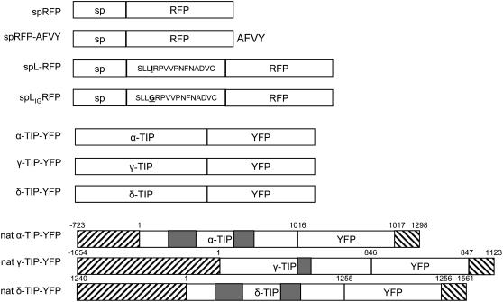
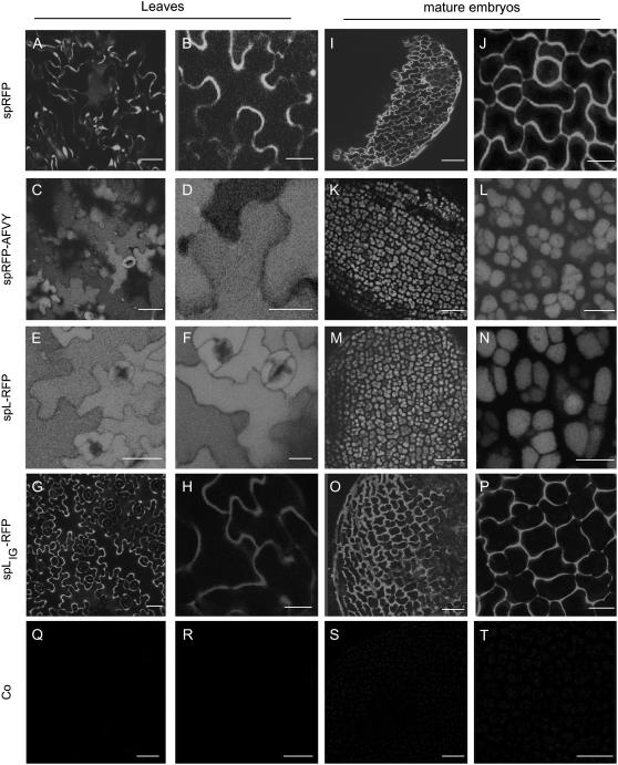
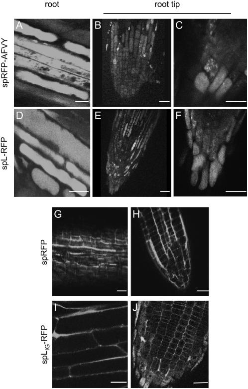
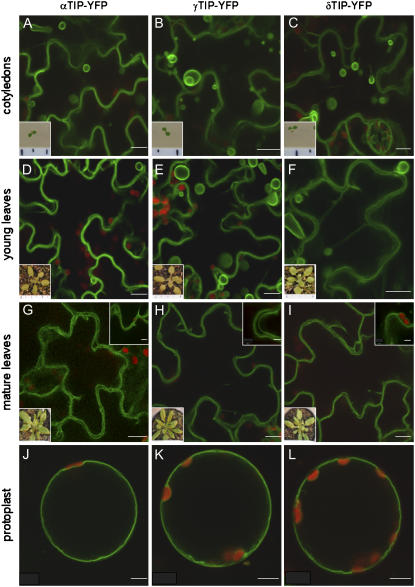
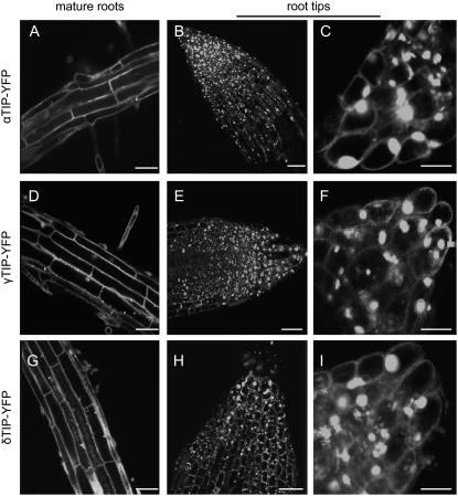
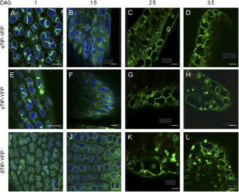
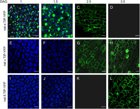
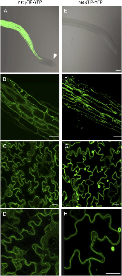
We generated fusions between three Arabidopsis (Arabidopsis thaliana) tonoplast intrinsic proteins (TIPs; alpha-, gamma-, and delta-TIP) and yellow fluorescent protein (YFP). We also produced soluble reporters consisting of the monomeric red fluorescent protein (RFP) and either the C-terminal vacuolar sorting signal of phaseolin or the sequence-specific sorting signal of proricin. In transgenic Arabidopsis leaves, mature roots, and root tips, all TIP fusions localized to the tonoplast of the central vacuole and both of the lumenal RFP reporters were found within TIP-delimited vacuoles. In embryos from developing, mature, and germinating seeds, all three TIPs localized to the tonoplast of protein storage vacuoles. To determine the temporal TIP expression patterns and to rule out mistargeting due to overexpression, we generated plants expressing YFP fused to the complete genomic sequences of the three TIP isoforms. In transgenic Arabidopsis, gamma-TIP expression was limited to vegetative tissues, but specifically excluded from root tips, whereas alpha-TIP was exclusively expressed during seed maturation. delta-TIP was expressed in vegetative tissues, but not root tips, at a later stage than gamma-TIP. Our findings indicate that, in the Arabidopsis tissues analyzed, two different vacuolar sorting signals target soluble proteins to a single vacuolar location. Moreover, TIP isoform distribution is tissue and development specific, rather than organelle specific.


Figures

Figure 1.
Figure 1.
Schematic representation of the constructs used in this study. Construct diagrams are not drawn to scale. sp, Signal peptide; L, proricin linker. Patterned boxes represent untranslated regions. Gray boxes represent introns.
Figure 2.
Figure 2.
Two vacuole-targeted RFP reporters localize to the same vacuole in Arabidopsis leaves and embryos. Leaves (A–H) and mature embryos (I–P) from transgenic plants expressing the indicated constructs or transformed with empty vector alone as a control (Co; Q–T) were analyzed by confocal microscopy. RFP was excited at 543 nm. All images shown were acquired using the same photomultiplier gain and offset settings. For each sample, two magnifications are shown. Scale bars: 40 μm (A, C, E, G, and Q, and I, K, M, O, and S) and 10 μm (B, D, F, H, and R, and J, L, N, P, and T).
Figure 3.
Figure 3.
Two vacuole-targeted RFP reporters localize to the same vacuole in Arabidopsis mature roots and root tips. Roots from transgenic plants expressing the indicated constructs were analyzed by confocal microscopy. Mature root sections and root tips are shown as indicated. RFP was excited at 543 nm. All images shown were acquired using the same photomultiplier gain and offset settings. For spRFP-AFVY and spL-RFP two magnifications are shown for the root tip. Scale bars: 10 μm.
Figure 4.
Figure 4.
All TIP-YFP fusions label the central vacuole of leaf cells. A to I, Leaves from transgenic Arabidopsis plants expressing the indicated reporter proteins at the stated growth stages were analyzed by confocal microscopy using YFP excited at 514 nm. YFP fluorescence is shown in green, chlorophyll autofluorescence in red. Scale bars: 10 μm. Insets (top right in G–I), Details of the TIP-YFP-labeled tonoplasts. Scale bars: 4 μm. Insets (bottom left in A–I), Stereomicrographs of the plants from which the leaves were taken. J to L, Protoplasts from leaves from the above transgenic plant lines were isolated and analyzed by confocal microscopy. Fluorescence is shown in green, chlorophyll autofluorescence in red. All images shown were acquired using the same photomultiplier gain and offset settings. Note that all three reporters localize to the tonoplast of the large central vacuole. Scale bar: 10 μm.
Figure 5.
Figure 5.
All TIP-YFP fusions label the same structures in roots. Mature roots and root tips from transgenic Arabidopsis plants expressing the indicated constructs were analyzed by confocal microscopy. A to C, α-TIP-YFP. D to F, γ-TIP-YFP. G to I, δ-TIP-YFP. Excitation was at 514 nm for YFP. Scale bars: 10 μm (A, B, D, E, G, and H) and 5 μm (C, F, and I).
Figure 6.
Figure 6.
TIP-YFP fusions label the PSV tonoplast during embryo germination. Embryos were dissected from seeds of Arabidopsis plants expressing the indicated constructs at the indicated time points after stratification. Embryos were analyzed by confocal microscopy. A to D, α-TIP-YFP. E to H, γ-TIP-YFP. I to L, δ-TIP-YFP. Excitation was at 405 nm for PSV autofluorescence (blue) and 514 nm for YFP (green). DAG, Days after germination. Scale bar: 10 μm.
Figure 7.
Figure 7.
YFP fusions to the TIP isoform genomic sequences highlight their temporal and tissue-specific regulation. Germinating seeds from the indicated Arabidopsis transgenic lines were imaged with a stereomicroscope at the indicated times. YFP was excited with a xenon lamp equipped with a GFP filter and detected with a digital camera. Chlorophyll autofluorescence appears red. All images were acquired using the same camera sensitivity and shutter speed. Insets show white-light images of the same samples. DAG, Days after germination.
Figure 8.
Figure 8.
α-TIP is replaced by γ- and δ-TIP during seed germination. Seeds from the indicated transgenic Arabidopsis lines were germinated and analyzed by confocal microscopy at the indicated time points. Excitation was at 405 nm for PSV autofluorescence (blue) and 514 nm for YFP (green). DAG, Days after germination. Scale bar: 10 μm.
Figure 9.
Figure 9.
Native δ- and γ-TIP are expressed in vegetative tissues but not root tips. Roots (A, B, E, and F) and leaves (C, D, G, and H) of transgenic Arabidopsis plants expressing the indicated constructs were analyzed by confocal microscopy. In A and E, YFP fluorescence is superimposed to the transmitted light image. Note that γ-TIP expression is restricted to the mature part of the root. Scale bars: 20 μm (A and E); 10 μm (B–D, F–H).

Comment in

  • Multiple vacuoles in plant cells.
    Rogers JC. Rogers JC. Plant Physiol. 2008 Mar;146(3):1024-5; author reply 1026-7. doi: 10.1104/pp.107.900248. Plant Physiol. 2008. PMID: 18316646 Free PMC article. No abstract available.

References

    1. Ahmed SU, Rojo E, Kovaleva V, Venkataraman S, Dombrowski KE, Matsuoka K, Raikhel NV (2000) The plant vacuolar sorting receptor AtELP is involved in transport of NH2-terminal propeptide-containing vacuolar proteins in Arabidopsis thaliana. J Cell Biol 149 1335–1344 - PMC - PubMed
    1. Batoko H, Zheng H-Q, Hawes C, Moore I (2000) A Rab1 GTPase is required for transport between the endoplasmic reticulum and Golgi apparatus and for normal Golgi movement in plants. Plant Cell 12 2201–2218 - PMC - PubMed
    1. Brown JC, Jolliffe NA, Frigerio L, Roberts LM (2003) Sequence-specific, Golgi-dependent targeting of the castor bean 2S albumin to the vacuole in tobacco protoplasts. Plant J 36 711–719 - PubMed
    1. Campbell RE, Tour O, Palmer AE, Steinbach PA, Baird GS, Zacharias DA, Tsien RY (2002) A monomeric red fluorescent protein. Proc Natl Acad Sci USA 99 7877–7882 - PMC - PubMed
    1. Carter C, Pan S, Zouhar J, Avila EL, Girke T, Raikhel NV (2004) The vegetative vacuole proteome of Arabidopsis thaliana reveals predicted and unexpected proteins. Plant Cell 16 3285–3303 - PMC - PubMed

Publication types

MeSH terms