. 2007 Oct 9;104(41):15988-93.
doi: 10.1073/pnas.0705062104. Epub 2007 Sep 28.

Collective migration of an epithelial monolayer in response to a model wound

Affiliations

Collective migration of an epithelial monolayer in response to a model wound

M Poujade et al. Proc Natl Acad Sci U S A. .

Abstract

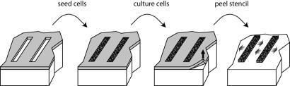
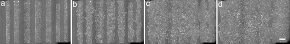
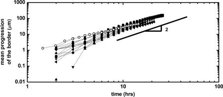
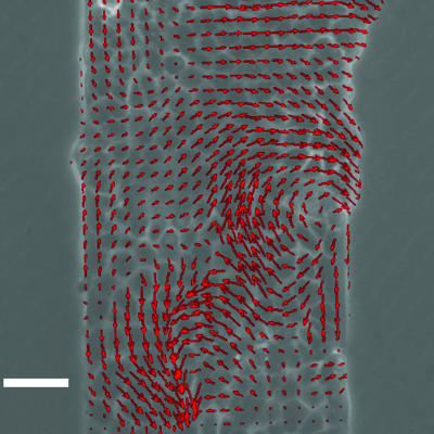
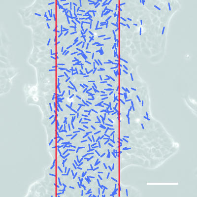
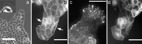
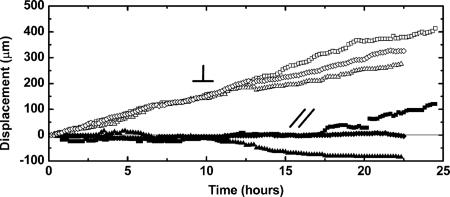
Using an original microfabrication-based technique, we experimentally study situations in which a virgin surface is presented to a confluent epithelium with no damage made to the cells. Although inspired by wound-healing experiments, the situation is markedly different from classical scratch wounding because it focuses on the influence of the free surface and uncouples it from the other possible contributions such as cell damage and/or permeabilization. Dealing with Madin-Darby canine kidney cells on various surfaces, we found that a sudden release of the available surface is sufficient to trigger collective motility. This migration is independent of the proliferation of the cells that mainly takes place on the fraction of the surface initially covered. We find that this motility is characterized by a duality between collective and individual behaviors. On the one hand, the velocity fields within the monolayer are very long range and involve many cells in a coordinated way. On the other hand, we have identified very active "leader cells" that precede a small cohort and destabilize the border by a fingering instability. The sides of the fingers reveal a pluricellular actin "belt" that may be at the origin of a mechanical signaling between the leader and the followers. Experiments performed with autocrine cells constitutively expressing hepatocyte growth factor (HGF) or in the presence of exogenous HGF show a higher average velocity of the border and no leader.


Conflict of interest statement

The authors declare no conflict of interest.

Figures

Fig. 1.
Fig. 1.
Principle of the experiments. The cells are cultured on an openware PDMS thin elastic film. When they reach confluence, this microstencil is removed to allow them to collectively migrate.
Fig. 2.
Fig. 2.
Sequence of micrographs showing the progression of several bands of different initial widths (a, t = 90 min; b, t = 13 h; c, t = 25 h; d, t = 37 h). The time t = 0 is taken at the removal of the stencil. Each image results from 18 acquisition fields stitched together. (Scale bar: 400 μm.)
Fig. 3.
Fig. 3.
Dynamic progression of the border of the epithelium. Several experiments performed in similar conditions are reported on the same graph. For the solid symbols, initial widths range from 150 μm to 400 μm. The open symbols represent an experiment with an initial width of 100 μm. The lines are guides for the eye.
Fig. 4.
Fig. 4.
Snapshot of the velocity field 4 h after removal of the stencil. This image was obtained by PIV (see text for details). The two vertices are an illustration of how coordinated the flows can be but are not a general feature. (Scale bar: 50 μm.)
Fig. 5.
Fig. 5.
Cumulative plot of the divisions over a time course of 16 h. Each blue line indicates the position and orientation of the division. The red lines indicate the initial position of the band. A vast majority (85%) of the divisions occur in the locus of this initial band with no particular orientation (see SI Fig. 9). (Scale bar: 100 μm.)
Fig. 6.
Fig. 6.
Micrographs of leader cells 18 h after stencil removal. In each image, a single leader drags a finger. (a) Phase contrast image of a finger preceded by a large leader cell. At the leading edge of this leader, there is a very active, ruffling lamellipodium (see Inset where the contrast has been enhanced on the same leader). (Scale bar: 100 μm.) (b) Fluorescence image of the actin cytoskeleton using Alexa 594-conjugated phalloidin staining. Particularly visible is the subcortical actin belt along the edges of the finger (arrows). (Scale bar: 50 μm.) (c) Immunofluorescence total internal reflection fluorescence image of the focal adhesions (vinculin labeling) showing that they are very developed at the leading edge of the leader. (Scale bar: 50 μm.) (d) Direct fluorescence image of the E-cadherin–GFP showing the cell–cell adhesions in particular between the leader and the followers. (Scale bar: 50 μm.)
Fig. 7.
Fig. 7.
Progression of the leaders for three independent fingers in two distinct experiments. Time t = 0 is taken at the beginning of the finger formation and not at the removal of the stencil. The symbol / / denotes the progression parallel to the initial edge (open symbols), the symbol ⊥ is used for displacements perpendicular to this initial edge (solid symbols). Leaders thus progress mostly perpendicularly to the initial wound edge at constant velocity.

Comment in

References

    1. Friedl P. Curr Opin Cell Biol. 2004;16:14–23. - PubMed
    1. Thiery J-P, Sleeman J. Nat Rev Mol Cell Biol. 2006;7:131–142. - PubMed
    1. Wood W, Jacinto A, Grose R, Gale J, Wilson C, Martin P. Nat Cell Biol. 2002;4:907–912. - PubMed
    1. Nobes CD, Hall A. J Cell Biol. 1999;144:1235–1244. - PMC - PubMed
    1. Martin P, Parkhurst S. Development (Cambridge, UK) 2004;131:3021–3034. - PubMed

Publication types

Substances

LinkOut - more resources