

DNA bending in the mycobacterial plasmid pAL5000 origin-RepB complex
- PMID: 17905972
- PMCID: PMC2168951
- DOI: 10.1128/JB.01155-07
DNA bending in the mycobacterial plasmid pAL5000 origin-RepB complex
Abstract
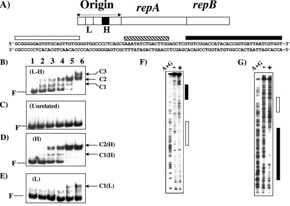
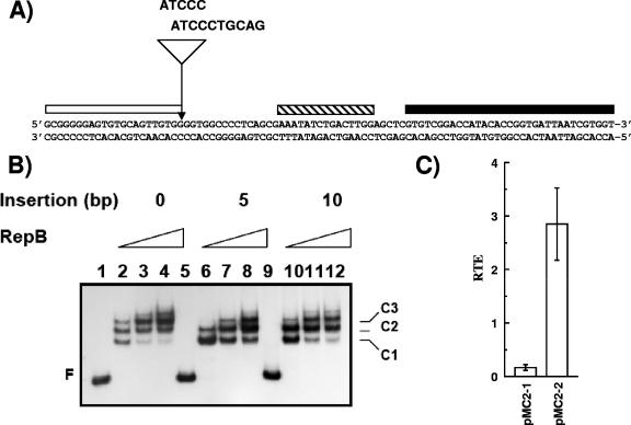
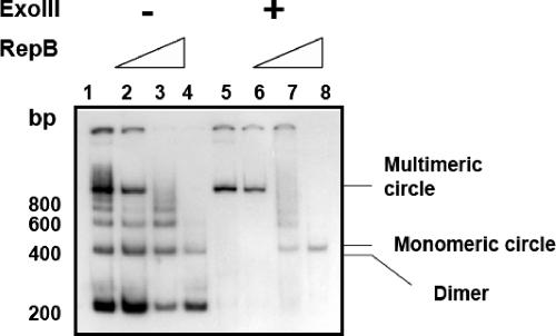
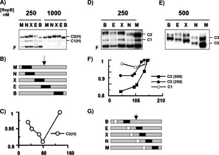
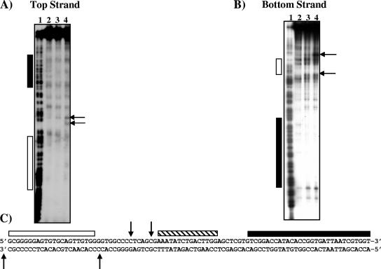
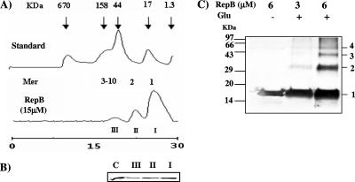
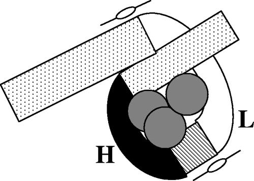
Plasmid pAL5000 represents a family of relatively newly discovered cryptic plasmids in gram-positive Actinomycetes bacteria. The replication regions of these plasmids comprise a bicistronic operon, repA-repB, encoding two replication proteins. Located upstream is a cis-acting element that functions as the origin of replication. It comprises an approximately 200-bp segment spanning two binding sites for the replication protein RepB, a low-affinity (L) site and a high-affinity (H) site separated by an approximately 40-bp spacer sequence. The trajectory of the DNA in the RepB-origin complex has been investigated, and it has been found that the origin undergoes significant bending movements upon RepB binding. RepB binding not only led to local bending effects but also caused a long-range polar curvature which affected the DNA sequences 3' to the H site. These movements appear to be essential for the in-phase alignment of the L and H sites that leads to the formation of a looped structure. A novel property of RepB unearthed in this study is its ability to form multimers. This property may be an important factor that determines the overall trajectory of the DNA in the RepB-origin complex. The results presented in this study suggest that the origins of replication of pAL5000 and related plasmids are highly flexible and that multimeric, RepB-like initiator proteins bind the origin and induce local deformations and long-range curvatures which are probably necessary for the proper functioning of the origin.
Figures
Similar articles
-
Origin binding activity of the Mycobacterial plasmid pAL5000 replication protein RepB is stimulated through interactions with host factors and coupled expression of repA.J Bacteriol. 2002 Apr;184(8):2204-14. doi: 10.1128/JB.184.8.2204-2214.2002. J Bacteriol. 2002. PMID: 11914352 Free PMC article.
-
Mutual interaction enables the mycobacterial plasmid pAL5000 origin binding protein RepB to recruit RepA, the plasmid replicase, to the origin.Microbiology (Reading). 2017 Apr;163(4):595-610. doi: 10.1099/mic.0.000447. Epub 2017 Apr 22. Microbiology (Reading). 2017. PMID: 28430099
-
Identification of promoter elements in mycobacteria: mutational analysis of a highly symmetric dual promoter directing the expression of replication genes of the Mycobacterium plasmid pAL5000.Nucleic Acids Res. 1999 Jan 15;27(2):396-402. doi: 10.1093/nar/27.2.396. Nucleic Acids Res. 1999. PMID: 9862957 Free PMC article.
-
Interactions between the RepB initiator protein of plasmid pMV158 and two distant DNA regions within the origin of replication.Nucleic Acids Res. 2007;35(4):1230-44. doi: 10.1093/nar/gkl1099. Epub 2007 Jan 31. Nucleic Acids Res. 2007. PMID: 17267412 Free PMC article.
-
The ABCs of plasmid replication and segregation.Nat Rev Microbiol. 2012 Nov;10(11):755-65. doi: 10.1038/nrmicro2882. Nat Rev Microbiol. 2012. PMID: 23070556 Review.
Cited by
-
Evolutionary link between the mycobacterial plasmid pAL5000 replication protein RepB and the extracytoplasmic function family of σ factors.J Bacteriol. 2012 Mar;194(6):1331-41. doi: 10.1128/JB.06218-11. Epub 2012 Jan 13. J Bacteriol. 2012. PMID: 22247504 Free PMC article.
-
RNase HI Depletion Strongly Potentiates Cell Killing by Rifampicin in Mycobacteria.Antimicrob Agents Chemother. 2022 Oct 18;66(10):e0209121. doi: 10.1128/aac.02091-21. Epub 2022 Sep 26. Antimicrob Agents Chemother. 2022. PMID: 36154174 Free PMC article.
-
Noncanonical SMC protein in Mycobacterium smegmatis restricts maintenance of Mycobacterium fortuitum plasmids.Proc Natl Acad Sci U S A. 2014 Sep 16;111(37):13264-71. doi: 10.1073/pnas.1414207111. Epub 2014 Sep 2. Proc Natl Acad Sci U S A. 2014. PMID: 25197070 Free PMC article.
References
-
- Aravind, L., V. Anantharam, S. Balaji, M. Mohan Babu, and L. M. Iyer. 2005. The many faces of the helix-turn-helix domain: transcription regulation and beyond. FEMS Microbiol. Rev. 29:231-262. - PubMed
-
- Chawla, M., and S. K. Das Gupta. 1999. Transposition-induced structural instability of Escherichia coli-mycobacteria shuttle vectors. Plasmid 41:135-140. - PubMed
Publication types
MeSH terms
Substances
LinkOut - more resources
Full Text Sources
Other Literature Sources
