. 2008 Aug 15;112(4):1329-37.
doi: 10.1182/blood-2007-08-107292. Epub 2007 Sep 28.

Anti-CS1 humanized monoclonal antibody HuLuc63 inhibits myeloma cell adhesion and induces antibody-dependent cellular cytotoxicity in the bone marrow milieu

Affiliations

Anti-CS1 humanized monoclonal antibody HuLuc63 inhibits myeloma cell adhesion and induces antibody-dependent cellular cytotoxicity in the bone marrow milieu

Yu-Tzu Tai et al. Blood. .

Abstract

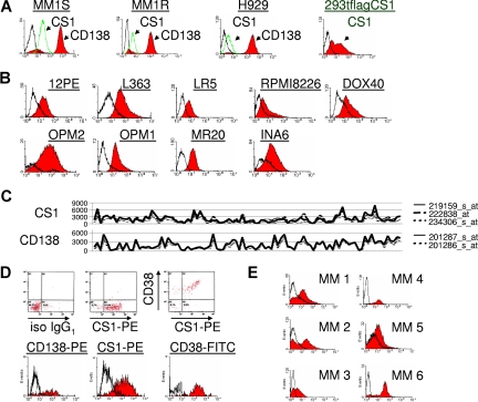
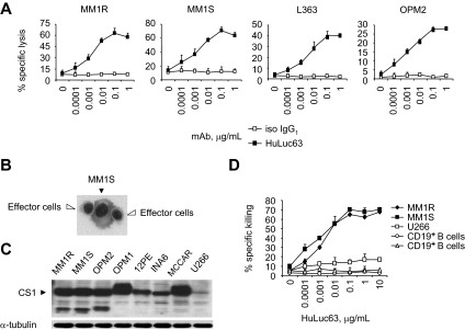
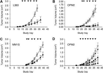
Currently, no approved monoclonal antibody (mAb) therapies exist for human multiple myeloma (MM). Here we characterized cell surface CS1 as a novel MM antigen and further investigated the potential therapeutic utility of HuLuc63, a humanized anti-CS1 mAb, for treating human MM. CS1 mRNA and protein was highly expressed in CD138-purified primary tumor cells from the majority of MM patients (more than 97%) with low levels of circulating CS1 detectable in MM patient sera, but not in healthy donors. CS1 was expressed at adhesion-promoting uropod membranes of polarized MM cells, and short interfering RNA (siRNA) targeted to CS1 inhibited MM cell adhesion to bone marrow stromal cells (BMSCs). HuLuc63 inhibited MM cell binding to BMSCs and induced antibody-dependent cellular cytotoxicity (ADCC) against MM cells in dose-dependent and CS1-specific manners. HuLuc63 triggered autologous ADCC against primary MM cells resistant to conventional or novel therapies, including bortezomib and HSP90 inhibitor; and pretreatment with conventional or novel anti-MM drugs markedly enhanced HuLuc63-induced MM cell lysis. Administration of HuLuc63 significantly induces tumor regression in multiple xenograft models of human MM. These results thus define the functional significance of CS1 in MM and provide the preclinical rationale for testing HuLuc63 in clinical trials, either alone or in combination.


Figures

Figure 1
Figure 1
CS1 expression in MM cell lines and patient MM cells. (A) MM1S, MM1R, and H929 MM cell lines were washed and immunostained with anti-CS1 (mouse clone 235614)-PE (green histogram), -CD138-PE (red histogram), or -isotype control IgG1 (iso IgG1)-PE. Open histograms to the left of each panel are for iso IgG1. (B) Immunostaining with a chimeric anti-CS1 ChLuc90 mAb (red histogram) or control human IgG (open histogram) was performed in 9 MM lines. (C) Total RNA isolated from CD138-purified tumor cells of each MM patient was subjected to microarray analysis using Affymetrix U133 Plus 2.0 array data. Three probes for CS1 and 2 probes for CD138 are indicated. (D) Expression of both CS1 (PE) and CD38 (FITC) in CD138-purified MM patient cells is shown (top). Open histograms represent iso IgG1, whereas solid histograms are for indicated antigens (bottom). (E) Immunostaining with ChLuc90 mAb or iso IgG1 in patient MM cells. Open and solid histograms represent iso IgG1 and CS1, respectively. 293tflagCS1 overexpressing CS1 was used as a positive control.
Figure 2
Figure 2
Serum level of circulating CS1 is detected only in myeloma patients. (A) CS1 ELISA-positive (MM6) or -negative (MM9) serum samples were immunoprecipitated (IP) with isotype control, HuLuc63, or ChLuc90 mAb covalently attached to Dynal Tosylactivated Dynabeads, followed by immunoblotting (IB) using anti-CS1 mAbs ChLuc90 (left) or 1G9 (right). (B) CS1 ELISA was done in serum samples from MM patients (n = 52) and healthy donors (n = 34). P was calculated by χ2 test from 2 × 2 contingency table (P < .001). N.D. indicates not detectable. (C) CS1 ELISA was performed in additional serum samples from newly diagnosed MM patients (n = 199). Patients with ISS I (n = 100) had significantly lower levels of CS1 than ISS II (n = 53) and III (n = 46; P = .006).
Figure 3
Figure 3
CS1 mediates MM cell adhesion to BMSC, which is blocked by HuLuc63. (A) MM lines and freshly isolated patient MM cells were incubated with 10 μg/mL of CS1-AF488 (green), washed, fixed, mounted, and viewed on a Zeiss microscope with a 40× objective. Images were processed using Adobe Photoshop Software version 7.0 (Adobe, San Jose, CA). CS1 is concentrated in uropods of polarized MM cells that promote adhesion. (B) CS1 (green) is colocalized with CD138 (red), exhibiting yellow staining in uropods of the majority of polarized L636 MM cells. (C) Lentiviruses expressing CS1 siRNA or control (cnt) siRNA were generated and used to infect MM1S cells. Immunoblotting using ChLuc90 mAb confirmed CS1 knockdown in clone 1. 293tflagCS1 expressing CS1 and 293t without CS1 expression served as controls. DAPI staining indicates nuclei of cells. (D) Calcein-AM labeled MM1S and MM1R cells, as well as CS1-null counterparts (MM1S CS1 siRNA and MM1R CS1 siRNA), were added to BMSC-coated 96-well plates, in the presence of iso IgG1 (□) or HuLuc63 (■; 0.1 μg/mL) for 4 hours. Unattached cells were washed and adherent cells were measured in a fluorescence plate reader. Shown is mean plus or minus SE of 3 independent experiments. A.U. indicates arbitrary unit. *P < .05. Error bars represent range of data. (E) Adhesion of MM1S and MM1R MM cells to BMSCs was assayed in the presence of HuLuc63 or iso IgG1. Shown is mean plus or minus SE of triplicate wells from one representative of 3 independent experiments. (F) HuLuc63 specifically inhibits patient MM cells (■ indicates MM1; ◆;; MM2) binding to BMSCs. (G) CS1 + CD138 + MM cells from 2 patients (MM 1 and MM 2) were incubated with HuLuc63 (0-100 μg/mL). (H) CS1 + MM cells from 2 patients were cultured with (■) or without (□) BMSCs, in the presence of HuLuc63 (0-100 μg/mL). Cell viability was determined by MTT assay. Shown is mean plus or minus SE of 3 independent experiments.
Figure 4
Figure 4
HuLuc63 triggers CS1-specific MM cell lysis through ADCC. (A) ADCC was performed by incubating calcein-AM–labeled target MM cells with human PBMC effector cells at an E/T ratio of 10:1, in the presence of various concentrations of HuLuc63 (■) or iso IgG1 (□). Percentage specific lysis was calculated, and data shown are representative of 3 experiments conducted with 3 different PBMC effector cell donors with similar results. HuLuc63 induces percent specific lysis of CS1-expressing MM lines in a dose-dependent manner. (B) Cytospin preparation of MM1S cells cultured with PBMC effector cells in the presence of HuLuc63 mAb (0.01 μg/mL) for 30 minutes was stained with Giemsa-Wright (Fisher Scientific, Springfield, NJ; original magnification ×200). (C) CS1 expression in 8 MM lines is determined by immunoblotting using anti-CS1 mAb. Only U266 has barely detectable CS1. (D) HuLuc63 does not stimulate dose-dependent ADCC against CS1 U266 MM line or CD19+ B cells from 2 healthy donors (open symbols), whereas it significantly induces lysis of CS1-expressing MM1R and MM1S target cells (solid symbols).
Figure 5
Figure 5
HuLuc63 mediates lysis of autologous MM cells resistant to conventional or novel therapies. (A) CD138-purified tumor cells from 9 patients with MM resistant or refractory to conventional therapies were incubated with autologous effector cells, in the presence of serial dilutions of HuLuc63 (■) or control iso IgG1 (□). Shown is mean plus or minus SE of triplicate wells. (B) CD138-purified tumor cells from 4 patients with MM resistant to either bortezomib or 17-AAG were incubated with autologous effector cells, in the presence of HuLuc63 (■) or iso IgG1 (□). Shown is mean plus or minus SE of triplicate wells. (C) PBMC effector cells were preincubated with lenalidomide (0.2 μM) followed by HuLuc63-mediated ADCC against MM1S and MM1R cells as well as primary MM patient cells. Shown is mean plus or minus SE of triplicate wells. (D) MM1R cells were pretreated overnight with U0126 (5 μM), Dex (25 nM), perifosine (5 μM), bortezomib (2 nM), or lenalidomide (0.05, 0.2 μM). HuLuc63-triggered ADCC against pretreated and control MM1R cells was assayed using PBMC effector cells from healthy donors (n = 3). HuLuc63, (■); iso IgG1 (□). *P < .05; **P < .01. (E) HuLuc63-mediated ADCC against MM1S and MM1R lines was performed in the presence or absence of BMSCs. *P < .05.
Figure 6
Figure 6
HuLuc63 exhibits antimyeloma activity in vivo and eradicates tumors in mice. (A-C) Mice with established myeloma xenograft tumors (average of approximately 100 mm3) were randomized into groups 16-21 days after inoculation and were then treated with either a humanized IgG1 control antibody (□) or HuLuc63 (▲). ▼ indicates the treatment days. Tumor growth results for individual mice are shown over a period of 40 days. Animals were taken off study once the tumors reached a size of greater than 2500 mm3. Group mean tumor volumes were significantly different between HuLuc63 and the control group in (A) the L363 model (P < .04 as of day 26); (B) the OPM2 model (P < .04 as of day 23); and (C) the MM1S model (P < .03 as of day 26). (D) Dose range finding study in the OPM2 model. Mice were randomized when tumors reached approximately 85 mm3 and were treated with control antibody at 10 mg/kg (□) or HuLuc63 at 0.1 mg/kg (♦), 0.5 mg/kg (●), 1 mg/kg (◇), and 10 mg/kg (▲). By day 23, all HuLuc63 groups reached significant difference from the control (with a minimum of P < .04), with the exception of the 0.1 mg/kg group, which was not significantly different from control throughout the study.

References

    1. Richardson PG, Sonneveld P, Schuster MW, et al. Bortezomib or high-dose dexamethasone for relapsed multiple myeloma. N Engl J Med. 2005;352:2487–2498. - PubMed
    1. Palumbo A, Bringhen S, Caravita T, et al. Oral melphalan and prednisone chemotherapy plus thalidomide compared with melphalan and prednisone alone in elderly patients with multiple myeloma: randomised controlled trial. Lancet. 2006;367:825–831. - PubMed
    1. Rajkumar SV, Blood E, Vesole D, Fonseca R, Greipp PR. Phase III clinical trial of thalidomide plus dexamethasone compared with dexamethasone alone in newly diagnosed multiple myeloma: a clinical trial coordinated by the Eastern Cooperative Oncology Group. J Clin Oncol. 2006;24:431–436. - PubMed
    1. Richardson PG, Blood E, Mitsiades CS, et al. A randomized phase 2 study of lenalidomide therapy for patients with relapsed or relapsed and refractory multiple myeloma. Blood. 2006;108:3458–3464. - PMC - PubMed
    1. Barlogie B, Tricot G, Anaissie E, et al. Thalidomide and hematopoietic-cell transplantation for multiple myeloma. N Engl J Med. 2006;354:1021–1030. - PubMed

Publication types