Beta-lactamase can function as a reporter of bacterial protein export during Mycobacterium tuberculosis infection of host cells
- PMID: 17906134
- PMCID: PMC2635098
- DOI: 10.1099/mic.0.2007/008516-0
Beta-lactamase can function as a reporter of bacterial protein export during Mycobacterium tuberculosis infection of host cells
Abstract
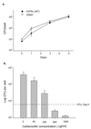
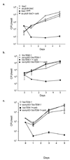
Mycobacterium tuberculosis is an intracellular pathogen that is able to avoid destruction by host immune defences. Exported proteins of M. tuberculosis, which include proteins localized to the bacterial surface or secreted into the extracellular environment, are ideally situated to interact with host factors. As a result, these proteins are attractive candidates for virulence factors, drug targets and vaccine components. Here we describe a beta-lactamase reporter system capable of identifying exported proteins of M. tuberculosis during growth in host cells. Because beta-lactams target bacterial cell-wall synthesis, beta-lactamases must be exported beyond the cytoplasm to protect against these drugs. When used in protein fusions, beta-lactamase can report on the subcellular location of another protein as measured by protection from beta-lactam antibiotics. Here we demonstrate that a truncated TEM-1 beta-lactamase lacking a signal sequence for export ('BlaTEM-1) can be used in this manner directly in a mutant strain of M. tuberculosis lacking the major beta-lactamase, BlaC. The 'BlaTEM-1 reporter conferred beta-lactam resistance when fused to both Sec and Tat export signal sequences. We further demonstrate that beta-lactamase fusion proteins report on protein export while M. tuberculosis is growing in THP-1 macrophage-like cells. This genetic system should facilitate the study of proteins exclusively exported in the host environment by intracellular M. tuberculosis.
Figures


Similar articles
-
Genome-wide identification of Mycobacterium tuberculosis exported proteins with roles in intracellular growth.J Bacteriol. 2011 Feb;193(4):854-61. doi: 10.1128/JB.01271-10. Epub 2010 Dec 10. J Bacteriol. 2011. PMID: 21148733 Free PMC article.
-
The twin-arginine translocation pathway of Mycobacterium smegmatis is functional and required for the export of mycobacterial beta-lactamases.J Bacteriol. 2005 Nov;187(22):7667-79. doi: 10.1128/JB.187.22.7667-7679.2005. J Bacteriol. 2005. PMID: 16267291 Free PMC article.
-
Identification of Mycobacterium tuberculosis signal sequences that direct the export of a leaderless beta-lactamase gene product in Escherichia coli.Microbiology (Reading). 1998 Jun;144 ( Pt 6):1619-1629. doi: 10.1099/00221287-144-6-1619. Microbiology (Reading). 1998. PMID: 9639933
-
The ins and outs of Mycobacterium tuberculosis protein export.Tuberculosis (Edinb). 2012 Mar;92(2):121-32. doi: 10.1016/j.tube.2011.11.005. Epub 2011 Dec 21. Tuberculosis (Edinb). 2012. PMID: 22192870 Free PMC article. Review.
-
Reappraising the use of β-lactams to treat tuberculosis.Expert Rev Anti Infect Ther. 2012 Sep;10(9):999-1006. doi: 10.1586/eri.12.96. Expert Rev Anti Infect Ther. 2012. PMID: 23106275 Free PMC article. Review.
Cited by
-
Identification of functional Tat signal sequences in Mycobacterium tuberculosis proteins.J Bacteriol. 2008 Oct;190(19):6428-38. doi: 10.1128/JB.00749-08. Epub 2008 Jul 25. J Bacteriol. 2008. PMID: 18658266 Free PMC article.
-
Protein export systems of Mycobacterium tuberculosis: novel targets for drug development?Future Microbiol. 2010 Oct;5(10):1581-97. doi: 10.2217/fmb.10.112. Future Microbiol. 2010. PMID: 21073315 Free PMC article. Review.
-
Protein export by the mycobacterial SecA2 system is determined by the preprotein mature domain.J Bacteriol. 2013 Feb;195(4):672-81. doi: 10.1128/JB.02032-12. Epub 2012 Nov 30. J Bacteriol. 2013. PMID: 23204463 Free PMC article.
-
Green Fluorescent Protein as a protein localization and topological reporter in mycobacteria.Tuberculosis (Edinb). 2017 Jul;105:13-17. doi: 10.1016/j.tube.2017.04.001. Epub 2017 Apr 10. Tuberculosis (Edinb). 2017. PMID: 28610783 Free PMC article.
-
Optimized APEX2 peroxidase-mediated proximity labeling in fast- and slow-growing mycobacteria.Methods Enzymol. 2022;664:267-289. doi: 10.1016/bs.mie.2021.11.021. Epub 2021 Dec 30. Methods Enzymol. 2022. PMID: 35331378 Free PMC article.
References
-
- Braunstein M, Espinosa BJ, Chan J, Belisle JT, Jacobs WR., Jr SecA2 functions in the secretion of superoxide dismutase A and in the virulence of Mycobacterium tuberculosis. Mol Microbiol. 2003;48:453–464. - PubMed
-
- Broome-Smith JK, Tadayyon M, Zhang Y. Beta-lactamase as a probe of membrane protein assembly and protein export. Mol Microbiol. 1990;4:1637–1644. - PubMed
Publication types
MeSH terms
Substances
Grants and funding
- T32 GM008581/GM/NIGMS NIH HHS/United States
- 5T32 AI07001-28/AI/NIAID NIH HHS/United States
- T32 AI007001/AI/NIAID NIH HHS/United States
- AI47311/AI/NIAID NIH HHS/United States
- 5-T32- GM008581/GM/NIGMS NIH HHS/United States
- R03 AI070928/AI/NIAID NIH HHS/United States
- 9P30 AI 50410-04/AI/NIAID NIH HHS/United States
- P30 AI050410/AI/NIAID NIH HHS/United States
- R01 AI054540/AI/NIAID NIH HHS/United States
- R01 AI047311/AI/NIAID NIH HHS/United States
- AI54540/AI/NIAID NIH HHS/United States
- AI070928/AI/NIAID NIH HHS/United States
LinkOut - more resources
Full Text Sources
Other Literature Sources
Research Materials
Miscellaneous

