

Cortical brain development in schizophrenia: insights from neuroimaging studies in childhood-onset schizophrenia
- PMID: 17906336
- PMCID: PMC2632387
- DOI: 10.1093/schbul/sbm103
Cortical brain development in schizophrenia: insights from neuroimaging studies in childhood-onset schizophrenia
Abstract
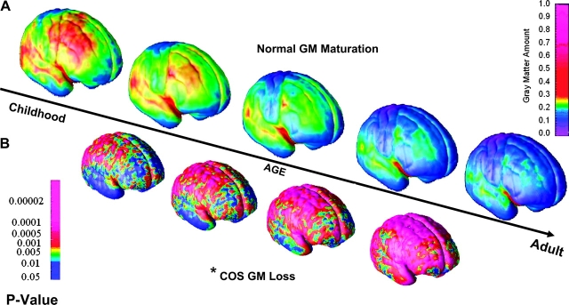
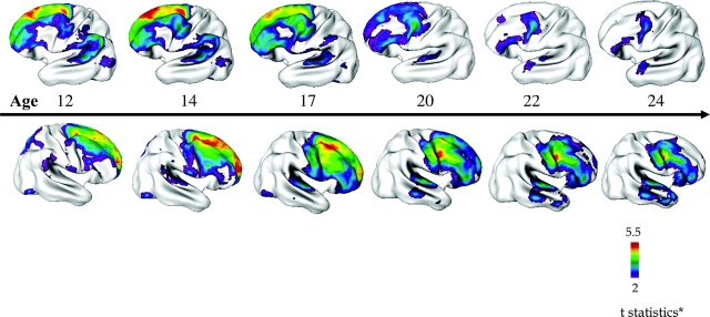
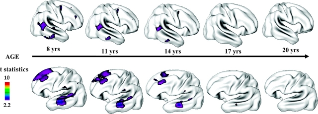
Childhood-onset schizophrenia (COS; defined as onset by age 12 years) is rare, difficult to diagnose, and represents a severe and chronic phenotype of the adult-onset illness. A study of childhood-onset psychoses has been ongoing at the National Institute of Mental Health (NIMH) since 1990, where children with COS and severe atypical psychoses (provisionally labeled "multidimensionally impaired" or MDI by the NIMH team) are studied prospectively along with all first-degree relatives. COS subjects have robust cortical gray matter (GM) loss during adolescence, which appears to be an exaggeration of the normal cortical GM developmental pattern and eventually mimics the pattern seen in adult-onset cases as the children become young adults. These cortical GM changes in COS are diagnostically specific and seemingly unrelated to the effects of medications. Furthermore, the cortical GM loss is also shared by healthy full siblings of COS probands suggesting a genetic influence on the abnormal brain development.
Figures
Similar articles
-
Comparison of progressive cortical gray matter loss in childhood-onset schizophrenia with that in childhood-onset atypical psychoses.Arch Gen Psychiatry. 2004 Jan;61(1):17-22. doi: 10.1001/archpsyc.61.1.17. Arch Gen Psychiatry. 2004. PMID: 14706940
-
Cortical brain development in nonpsychotic siblings of patients with childhood-onset schizophrenia.Arch Gen Psychiatry. 2007 Jul;64(7):772-80. doi: 10.1001/archpsyc.64.7.772. Arch Gen Psychiatry. 2007. PMID: 17606811
-
Childhood onset schizophrenia: cortical brain abnormalities as young adults.J Child Psychol Psychiatry. 2006 Oct;47(10):1003-12. doi: 10.1111/j.1469-7610.2006.01658.x. J Child Psychol Psychiatry. 2006. PMID: 17073979
-
Neuroimaging findings from childhood onset schizophrenia patients and their non-psychotic siblings.Schizophr Res. 2016 Jun;173(3):124-131. doi: 10.1016/j.schres.2015.03.003. Epub 2015 Mar 26. Schizophr Res. 2016. PMID: 25819937 Free PMC article. Review.
-
Imaging normal and abnormal brain development: new perspectives for child psychiatry.Aust N Z J Psychiatry. 2001 Jun;35(3):272-81. doi: 10.1046/j.1440-1614.2001.00900.x. Aust N Z J Psychiatry. 2001. PMID: 11437799 Review.
Cited by
-
Delayed white matter growth trajectory in young nonpsychotic siblings of patients with childhood-onset schizophrenia.Arch Gen Psychiatry. 2012 Sep;69(9):875-84. doi: 10.1001/archgenpsychiatry.2011.2084. Arch Gen Psychiatry. 2012. PMID: 22945617 Free PMC article.
-
Progress in iPSC-Based Modeling of Psychiatric Disorders.Int J Mol Sci. 2019 Oct 2;20(19):4896. doi: 10.3390/ijms20194896. Int J Mol Sci. 2019. PMID: 31581684 Free PMC article. Review.
-
Gray matter alterations in schizophrenia high-risk youth and early-onset schizophrenia: a review of structural MRI findings.Child Adolesc Psychiatr Clin N Am. 2013 Oct;22(4):689-714. doi: 10.1016/j.chc.2013.06.003. Epub 2013 Jul 23. Child Adolesc Psychiatr Clin N Am. 2013. PMID: 24012081 Free PMC article. Review.
-
Editorial: research progress in early-onset schizophrenia.Schizophr Bull. 2008 Jan;34(1):15-7. doi: 10.1093/schbul/sbm123. Epub 2007 Nov 28. Schizophr Bull. 2008. PMID: 18048380 Free PMC article.
-
Dendritic Spine Initiation in Brain Development, Learning and Diseases and Impact of BAR-Domain Proteins.Cells. 2021 Sep 12;10(9):2392. doi: 10.3390/cells10092392. Cells. 2021. PMID: 34572042 Free PMC article. Review.
References
-
- Childs B, Scriver CR. Age at onset and causes of disease. Perspect Biol Med. 1986;29(Pt 1):437–460. - PubMed
-
- Nicolson R, Rapoport JL. Childhood-onset schizophrenia: rare but worth studying. Biol Psychiatry. 1999;46:1418–1428. - PubMed
-
- Kraepelin E. Dementia Praecox and Paraphrenia. 1919 ed. Huntington, NY: Robert E Krieger; 1919.
-
- Volkmar FR. Childhood and adolescent psychosis: a review of the past 10 years. J Am Acad Child Adolesc Psychiatry. 1996;35:843–851. - PubMed
-
- Kolvin I. Studies in the childhood psychoses. I. Diagnostic criteria and classification. Br J Psychiatry. 1971;118:381–384. - PubMed
Publication types
MeSH terms
Grants and funding
LinkOut - more resources
Full Text Sources
Medical
