. 2007 Nov;10(11):1483-91.
doi: 10.1038/nn1967. Epub 2007 Sep 30.

Attention alters spatial integration in macaque V1 in an eccentricity-dependent manner

Affiliations

Attention alters spatial integration in macaque V1 in an eccentricity-dependent manner

Mark Roberts et al. Nat Neurosci. 2007 Nov.

Abstract

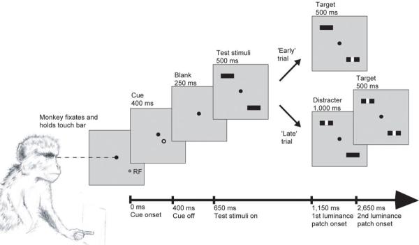
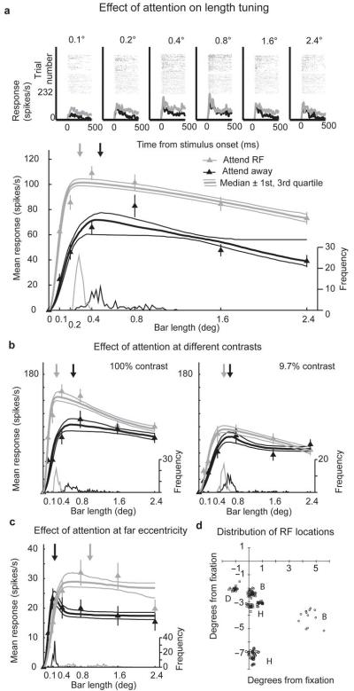
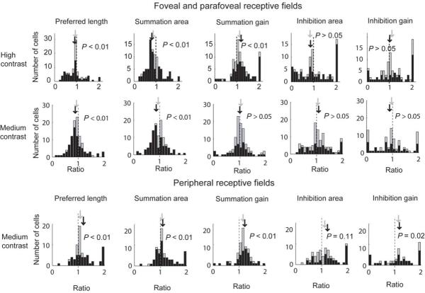
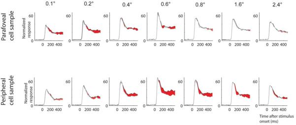
Attention can selectively enhance neuronal responses and exclude external noise, but the neuronal computations that underlie these effects remain unknown. At the neuronal level, noise exclusion might result in altered spatial integration properties. We tested this proposal by recording neuronal activity and length tuning in neurons of the primary visual cortex of the macaque when attention was directed toward or away from stimuli presented in each neuron's classical receptive field. For cells with central-parafoveal receptive fields, attention reduced spatial integration, as demonstrated by a reduction in preferred stimulus length and in the size of the spatial summation area. Conversely, in cells that represented more peripheral locations, attention increased spatial integration by increasing the cell's summation area. This previously unknown dichotomy between central and peripheral vision could support accurate analysis of attended foveal objects and target selection for impending eye movements to peripheral objects.


Figures

Figure 1
Figure 1
Representation of the main experimental task. The timing of events relative to the start of the trial is marked along the bottom axis. Presentation time is given above each frame. The monkey initiated the trial by fixating centrally (filled circle) and holding a touch-bar. At the start of the trial a cue (open circle) indicated the location to which the monkey should attend, while the cue itself was spatially and temporally separated from the stimulus and thus the attended location. In the current example the cue directs attention towards the RF of the neuron under study. Test stimuli were two identical bars, one presented at the RF of the neuron under study, the other in the opposite hemifield. The monkey’s task was to detect the presentation of a 0.1°×0.1° luminance patch occurring centrally on the cued bar. This occurred at either 500 ms or 1,500 ms after the onset of the test stimuli. In an ‘early’ trial the first luminance patch was presented on the cued stimulus (‘target’). In a ‘late’ trial the first luminance patch was presented on the un-cued test stimulus (‘distracter’) and the second patch occurred on the cued stimulus. The monkey had 500 ms to release the touch bar following the presentation of a target, in order to receive a juice reward.
Figure 2
Figure 2
Effect of attention on length tuning for individual cells. a) Upper plots show raster plots and histograms of single cell responses for each bar length. Data in grey show the attend-RF condition, data in black show the attend-away condition. Lower plot shows the effect of attention on length tuning. Triangles show mean response at each bar length, error bars s.e.m. Bold line fitted to the data shows the median DOG model fit from the bootstrap procedure, flanking upper and lower narrow lines show the 75th and 25th percentile fits. Curves at the base of each plot show the distribution of preferred lengths taken from 100 iterations of the bootstrap procedure. The frequency values of the histogram are shown on the rightward y-axis. The median preferred length is marked with the downwards-pointing arrow. Grey triangles, lines and arrows show data from the attend-RF condition; black triangles, lines and arrows show data from the attend-away condition. Error bars show s.e.m. This cell was recorded using high contrast stimuli. b) Example cell showing the effect of attention on length tuning at both high and medium contrast. Data are shown in the same format as in part a, the cell was recorded from monkey D. c) Example cell with a receptive field eccentricity of ~6° (monkey B). d) Distribution of RF locations. Each point marks the location of a RF. Capital letters next to RF clusters indicate which monkey the respective cells were recorded from (e.g. B:=monkey B).
Figure 3
Figure 3
Effect of attention on preferred length and on DOG summation area and summation gain for high (top row) and medium contrast data (middle row) recorded at ~ 2-3° eccentricity location, as well as for medium contrast data recorded at the ~6-7° eccentricity location (bottom row). Values below 1 (vertical dashed line) indicate that the parameter of interest was reduced in the attend-RF condition. Light shaded histograms show the distribution of ratios across the population of cells. Dark shaded histograms show the distribution of ratios across cells for which the parameter of interest was significantly different between the two conditions, assessed by bootstrapping. The median ratio for the whole population and the population of significant cells is marked with downwards pointing arrows, shaded grey and black respectively. P values give the significance of changes in the parameter of interest with attention (whole population signed rank test Ho median ratio=1).
Figure 4
Figure 4
Population response as a function of bar length. Upper row of histograms: Response from cells recorded in monkey B (n = 17) and H (n = 56) at medium contrast from parafoveal sites. Lower row of histograms: Response from cells recorded in monkey B (n = 22) and H (n = 47) at medium contrast from peripheral sites. Black curves: response for the attend RF condition, grey curves: response for the attend away condition. Red shaded areas: time periods during which response differences were significant for the population (P < 0.01, rank sum test). Response normalization was performed for each cell across all conditions initially. Population responses were calculated from these individually normalized responses. Population histogram for bar-length 0.6 deg is from monkey H only, as recordings in monkey B were restricted to 6 different bar-lengths. Data from monkey B and H are included as both contributed to our parafoveal and peripheral cell sample.
Figure 5
Figure 5
Quantitative comparison of attentional modulation as a function of bar-length and recording location. Attentional modulation was assessed by calculating a receiver operating characteristic (ROC) as a function of bar-length for each cell from monkey B and monkey H (recordings from medium contrast experiments) for parafoveal sites (black solid curve) and for peripheral sites (black dotted curve). ROC values differed significantly as a function of bar-length for both recording sites (P < 0.001, ANOVA on ranks). Error bars show s.e.m. Data for bar-length 0.6 deg are from monkey H only, as recordings in monkey B were restricted to 6 different bar-lengths. Data from monkey D are not included, as he did not contribute to data from peripheral sites.

References

    1. Haenny PE, Schiller PH. State dependent activity in monkey visual cortex. I Single cell activity in V1 and V4 on visual tasks. Exp.Brain Res. 1988;69:225–244. - PubMed
    1. Luck SJ, Chelazzi L, Hillyard SA, Desimone R. Neural mechanisms of spatial selective attention in areas V1, V2, and V4 of macaque visual cortex. J Neurophysiol. 1997;77:24–42. - PubMed
    1. Roelfsema PR, Lamme VA, Spekreijse H. Object-based attention in the primary visual cortex of the macaque monkey. Nature. 1998;395:376–81. - PubMed
    1. Spitzer H, Desimone R, Moran J. Increased Attention Enhances Both Behavioral and Neuronal Performance. Science. 1988;240:338–340. - PubMed
    1. Treue S, Maunsell JHR. Attentional modulation of visual motion processing in cortical areas MT and MST. Nature. 1996;382:539–541. - PubMed

Publication types