

Histological changes at an endosonography-guided biliary drainage site: a case report
- PMID: 17907298
- PMCID: PMC4171289
- DOI: 10.3748/wjg.v13.i41.5512
Histological changes at an endosonography-guided biliary drainage site: a case report
Abstract
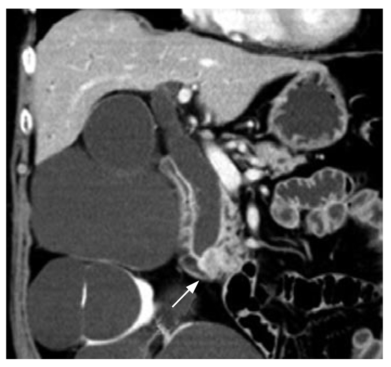
Endosonography-guided biliary drainage (ESBD) is a new method enabling internal drainage of an obstructed bile duct. However, the histological conditions associated with fistula development via the duodenum to the bile duct have not been reported. We performed ESBD 14 d preoperatively in a patient with an ampullary carcinoma and histologically confirmed changes in and around the fistula. The female patient developed no complications relevant to ESBD. Levels of serum bilirubin and hepatobiliary enzymes declined quickly, and pancreatoduodenectomy was carried out uneventfully. The resected specimen was sliced and stained with hematoxylin-eosin. Histological evaluation of the puncture site in the duodenum and bile-duct wall, and the sinus tract revealed no hematoma, bile leakage, or abscess in or around the sinus tract. Little sign of granulation, fibrosis, and inflammatory cell infiltration was observed. Although further large-scale confirmatory studies are needed, the findings here may encourage more active use of ESBD as a substitute for percutaneous transhepatic drainage in cases with failed/difficult endoscopic biliary stenting.
Figures


References
-
- Harada N, Kouzu T, Arima M, Asano T, Kikuchi T, Isono K. Endoscopic ultrasound-guided pancreatography: a case report. Endoscopy. 1995;27:612–615. - PubMed
-
- Wiersema MJ, Sandusky D, Carr R, Wiersema LM, Erdel WC, Frederick PK. Endosonography-guided cholangiopancreatography. Gastrointest Endosc. 1996;43:102–106. - PubMed
-
- Giovannini M, Moutardier V, Pesenti C, Bories E, Lelong B, Delpero JR. Endoscopic ultrasound-guided bilioduodenal anastomosis: a new technique for biliary drainage. Endoscopy. 2001;33:898–900. - PubMed
-
- Burmester E, Niehaus J, Leineweber T, Huetteroth T. EUS-cholangio-drainage of the bile duct: report of 4 cases. Gastrointest Endosc. 2003;57:246–251. - PubMed
-
- Mallery S, Matlock J, Freeman ML. EUS-guided rendezvous drainage of obstructed biliary and pancreatic ducts: Report of 6 cases. Gastrointest Endosc. 2004;59:100–107. - PubMed
Publication types
MeSH terms
LinkOut - more resources
Full Text Sources
