

Case Reports
doi: 10.3748/wjg.v13.i41.5525.
Gallstone spillage caused by spontaneously perforated hemorrhagic cholecystitis
Affiliations
- PMID: 17907301
- PMCID: PMC4171292
- DOI: 10.3748/wjg.v13.i41.5525
Item in Clipboard
Case Reports
Gallstone spillage caused by spontaneously perforated hemorrhagic cholecystitis
World J Gastroenterol.
.
Abstract
There are occasional incidences of gallstone spillage during laparoscopic cholecystectomy, and there have been frequent reports on such a topic in the literature. To the best of our knowledge, however, there have been no reports about spilled stones caused by spontaneously perforated hemorrhagic cholecystitis. Here, we report the radiologic findings of spilled stones caused by spontaneously perforated hemorrhagic cholecystitis in a 55-year-old man.
Figures
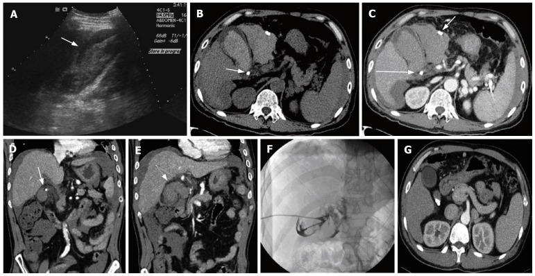
A 55-year-old man with right upper quadrant pain. US images (A) demonstrate heterogeneous, highly echogenic material, both within and outside the gallbladder lumen (arrows), with a positive sonographic Murphy's sign. Non-contrast (B) and contrast-enhanced (C) transverse CT images show high-attenuation (46-61HU) material, both in the gallbladder lumen and pericholecystic space. One stone is seen in the cystic duct (long arrow) and calcified material (with the same appearance as the cystic duct stone) is seen in the fluid collected (short arrow) around the gallbladder. Contrast-enhanced coronal CT images (D, E) show well the impacted cystic-duct stone (arrow), and the mucosal defect with continuation of hemorrhage (dotted arrow). PTGBD (F) with cholecystography demonstrates contrast leakage from the gallbladder. Contrast-enhanced transverse CT images (G) taken 2 mo before the current attack show multiple stones in the gallbladder neck without complications.
References
-
- Ahmad SA, Schuricht AL, Azurin DJ, Arroyo LR, Paskin DL, Bar AH, Kirkland ML. Complications of laparoscopic cholecystectomy: the experience of a university-affiliated teaching hospital. J Laparoendosc Adv Surg Tech A. 1997;7:29–35. - PubMed
-
- Woodfield JC, Rodgers M, Windsor JA. Peritoneal gallstones following laparoscopic cholecystectomy: incidence, complications, and management. Surg Endosc. 2004;18:1200–1207. - PubMed
-
- Hui TT, Giurgiu DI, Margulies DR, Takagi S, Iida A, Phillips EH. Iatrogenic gallbladder perforation during laparoscopic cholecystectomy: etiology and sequelae. Am Surg. 1999;65:944–948. - PubMed
Publication types
MeSH terms
LinkOut - more resources
Full Text Sources
Medical
