Carcinosarcoma of the stomach: a case report and review of the literature
- PMID: 17907304
- PMCID: PMC4171295
- DOI: 10.3748/wjg.v13.i41.5533
Carcinosarcoma of the stomach: a case report and review of the literature
Abstract
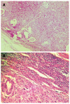
Carcinosarcomas are rare, malignant, biphasic tumors. We report the case of a 62-year-old man with gastric carcinosarcoma, along with its clinical, macroscopic and histopathological features. Macroscopically, a specimen of deformed stomach was obtained that measured 200 mm x 150 mm x 100 mm. A 150 mm x 100 mm x 50 mm exophytic tumoral mass (Borrmann type I) was found, which involved the posterior wall from the cardia to the antrum. Histopathologically, a mixed type of malignancy was revealed: an adenocarcinoma with intestinal metaplasia, with interposed fascicles of fusiform atypical cells and numerous large, rounded and oval cells. The tumor showed positive histochemistry for cytokeratin 18, epithelial membrane antigen, carcinoembryonic antigen, chromogranin A and vimentin. Liver metastases were diagnosed 8 mo postoperatively, and the patient died 4 mo later. A review of the available literature is also presented.
Figures


References
-
- Kanamoto A, Nakanishi Y, Ochiai A, Shimoda T, Yamaguchi H, Tachimori Y, Kato H, Watanabe H. A case of small polypoid esophageal carcinoma with multidirectional differentiation, including neuroendocrine, squamous, ciliated glandular, and sarcomatous components. Arch Pathol Lab Med. 2000;124:1685–1687. - PubMed
-
- Yamazaki K. A gastric carcinosarcoma with neuroendocrine cell differentiation and undifferentiated spindle-shaped sarcoma component possibly progressing from the conventional tubular adenocarcinoma; an immunohistochemical and ultrastructural study. Virchows Arch. 2003;442:77–81. - PubMed
-
- Insabato L, Di Vizio D, Ciancia G, Pettinato G, Tornillo L, Terracciano L. Malignant gastrointestinal leiomyosarcoma and gastrointestinal stromal tumor with prominent osteoclast-like giant cells. Arch Pathol Lab Med. 2004;128:440–443. - PubMed
-
- Kayaselcuk F, Tuncer I, Toyganözü Y, Bal N, Ozgür G. Carcinosarcoma of the stomach. Pathol Oncol Res. 2002;8:275–277. - PubMed
-
- Teramachi K, Kanomata N, Hasebe T, Ishii G, Sugito M, Ochiai A. Carcinosarcoma (pure endocrine cell carcinoma with sarcoma components) of the stomach. Pathol Int. 2003;53:552–556. - PubMed
Publication types
MeSH terms
Substances
LinkOut - more resources
Full Text Sources
Medical
Research Materials
Miscellaneous

