Subperiosteal chondromyxoid fibroma: a report of two cases
- PMID: 17907440
- PMCID: PMC2150655
Subperiosteal chondromyxoid fibroma: a report of two cases
Abstract
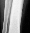
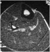
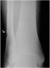
Chondromyxoid fibroma is a rare cartilage tumor that represents less than 1% of all bone tumors. When in a long bone, it is usually an intramedullary lesion that is eccentrically located in the metaphyseal region. Chondromyxoid fibroma may also have unusual presentations. These include intracortical lesions and subperiosteal lesions. There have been 14 reported cases of intracortical chondromyxoid fibroma, but there have been only four reports of subperiosteal lesions. A subperiosteal location, therefore, is extremely rare for a chondromyxoid fibroma. We present two new cases of subperiosteal chondromyxoid fibroma. Given its rarity, chondromyxoid fibroma is often not in the differential diagnosis of a painful, subperiosteal scalloped lesion in a long bone. Other entities such as periosteal chondroma, periosteal myxoma, subperiosteal ganglion cyst, or subperiosteal osteoid osteoma are more likely to be considered. Our cases illustrates that subperiosteal chondromyxoid fibroma, although rare, should be included in the differential diagnosis of a painful, radiographically inactive lytic lesion on the surface of a long bone.
Conflict of interest statement
Each author certifies that he has no commercial associations (e.g., consultancies, stock ownership, equity interest, patent/licensing arrangements, etc.) that might pose a conflict of interest in connection with the submitted article
Each author certifies that his institution has approved the reporting of this case series, that all investigations were conducted in conformity with ethical principles of research, and that informed consent was obtained.
Figures


Similar articles
-
Intracortical chondromyxoid fibroma of the tibia.Musculoskelet Surg. 2013 Aug;97(2):177-81. doi: 10.1007/s12306-011-0162-3. Epub 2011 Aug 4. Musculoskelet Surg. 2013. PMID: 21814765
-
Surface chondromyxoid fibroma of the distal ulna: unusual tumor, site, and age.Skeletal Radiol. 2014 Feb;43(2):243-6. doi: 10.1007/s00256-013-1720-6. Epub 2013 Sep 21. Skeletal Radiol. 2014. PMID: 24057439
-
Non specific magnetic resonance features of chondromyxoid fibroma of the iliac bone.J BUON. 2007 Jul-Sep;12(3):407-9. J BUON. 2007. PMID: 17918298
-
Chondromyxoid fibroma of the calcaneus: two case reports and literature review.J Foot Ankle Surg. 2013 Sep-Oct;52(5):643-9. doi: 10.1053/j.jfas.2013.02.014. Epub 2013 Apr 13. J Foot Ankle Surg. 2013. PMID: 23590809 Review.
-
Cytopathology of chondromyxoid fibroma: a case series and review of the literature.J Am Soc Cytopathol. 2021 Jul-Aug;10(4):366-381. doi: 10.1016/j.jasc.2021.04.001. Epub 2021 Apr 7. J Am Soc Cytopathol. 2021. PMID: 33958292 Review.
Cited by
-
Osteoid osteoma and osteoid osteoma-mimicking lesions: biopsy findings, distinctive MDCT features and treatment by radiofrequency ablation.Eur Radiol. 2010 Oct;20(10):2439-46. doi: 10.1007/s00330-010-1811-x. Epub 2010 May 15. Eur Radiol. 2010. PMID: 20467872
-
A Case Report of Chondromyxoid Fibroma of the Neck of Femur, Intracapsular Location.J Orthop Case Rep. 2021;11(1):79-81. doi: 10.13107/jocr.2021.v11.i01.1972. J Orthop Case Rep. 2021. PMID: 34141648 Free PMC article.
-
Chondromyxoid Fibroma of the Calcaneus: A Rare Case Report.Cureus. 2022 Feb 6;14(2):e21950. doi: 10.7759/cureus.21950. eCollection 2022 Feb. Cureus. 2022. PMID: 35282516 Free PMC article.
-
Juxtacortical chondromyxoid fibroma in the small bones: two cases with unusual location and a literature review.J Pathol Transl Med. 2022 May;56(3):157-160. doi: 10.4132/jptm.2021.12.15. Epub 2022 Jan 21. J Pathol Transl Med. 2022. PMID: 35051327 Free PMC article.
-
Huge chondromyxoid fibroma of proximal third tibia masquerading as an aneurysmal bone cyst: A rare case report.South Asian J Cancer. 2013 Jan;2(1):13. doi: 10.4103/2278-330X.105875. South Asian J Cancer. 2013. PMID: 24455535 Free PMC article. No abstract available.
References
-
- Unni KK. Dahlin's Bone Tumors: General Aspects and Dataon1 1,087 Cases. fifth. Philadelphia: Lippincott-Raven; 1996. p. 59.
-
- Jaffe HL. Tumors and Tumorous Conditions of the Bones and Joints. Philadelphia: Lea and Febiger; 1958. p. 205.
-
- Ralph LL. Chondromyxoid fibroma of bone. J Bone Joint Surg Br. 1962;44-B:7–24. - PubMed
-
- Murray RD, Jacobson HG. The Radiology of Skeletal Disorders. Vol. 1. Baltimore: Williams and Wilkins; 1971. p. 402.
-
- Greenfield GB. Radiology of Bone Diseases. second. Philadelphia: Lippincott; 1975. pp. 465–467.
Publication types
MeSH terms
LinkOut - more resources
Full Text Sources
Medical
