

doi: 10.1371/journal.ppat.0030113.
Botulinum neurotoxin heavy chain belt as an intramolecular chaperone for the light chain
Affiliations
- PMID: 17907800
- PMCID: PMC1994969
- DOI: 10.1371/journal.ppat.0030113
Item in Clipboard
Botulinum neurotoxin heavy chain belt as an intramolecular chaperone for the light chain
PLoS Pathog.
.
No abstract available
Conflict of interest statement
Figures

(A) Structure of BoNT/A. The Cα backbone of the LC (left) is represented as cyan ribbons; the purple sphere highlights the catalytic Zn2+ at the protease active site. The HC belt segment encompassing residues 492–545 is displayed in magenta and the 449–491 region in gold. The HC is depicted in blue, in which the helical module (middle) is the translocation domain, and the two sub-domains consisting primarily of β-strands constitute the receptor-binding domain (right). (B) Structure of BoNT/A-LC in complex with the sn2 segment of SNAP-25 [16]. The Cα backbone of the LC is represented as cyan ribbons and its molecular surface in transparent grey. The sn2 segment is depicted in red and the catalytic Zn2+ at the active site as a purple sphere. (C) Superposition of the structures of the sn2 segment in complex with the LC/A, the HC belt of BoNT/A, and the HC belt of BoNT/B. LC removed for display. For the superposition, the backbone atoms of the LCs were used for the best fit between the structures. sn2 segment depicted in red; overlay of the Cα backbone of BoNT/A [4] and BoNT/B [6] belts represented as magenta and lime ribbons. Spheres represent the catalytic Zn2+ at the active site of BoNT/A (red and magenta) and BoNT/B (lime). All images were rendered with YASARA [40].
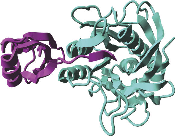
The Cα backbone of subtilisin is represented as cyan ribbons and that of the pro-domain in magenta. Note that the C-terminus of the pro-domain is lodged in a crevice at the protease active site; the tip of the β-strand highlights Y77 at the active site. Image rendered using subtilisin BPN' prosegment complexed with a mutant subtilisin BPN' [31,32] with YASARA [40].
References
-
- Arnon SS, Schechter R, Inglesby TV, Henderson DA, Bartlett JG, et al. Botulinum toxin as a biological weapon: Medical and public health management. JAMA. 2001;285:1059–1070. - PubMed
-
- Schiavo G, Matteoli M, Montecucco C. Neurotoxins affecting neuroexocytosis. Physiol Rev. 2000;80:717–766. - PubMed
-
- Lacy DB, Tepp W, Cohen AC, DasGupta BR, Stevens RC. Crystal structure of botulinum neurotoxin type A and implications for toxicity. Nat Struct Biol. 1998;5:898–902. - PubMed
-
- Lacy DB, Stevens RC. Sequence homology and structural analysis of the clostridial neurotoxins. J Mol Biol. 1999;291:1091–1104. - PubMed
Publication types
MeSH terms
Substances
Grants and funding
LinkOut - more resources
Full Text Sources
Other Literature Sources
