. 2007 Sep 7;3(9):1281-90.
doi: 10.1371/journal.ppat.0030134.

Apoptotic killing of HIV-1-infected macrophages is subverted by the viral envelope glycoprotein

Affiliations

Apoptotic killing of HIV-1-infected macrophages is subverted by the viral envelope glycoprotein

Simon Swingler et al. PLoS Pathog. .

Abstract

Viruses have evolved strategies to protect infected cells from apoptotic clearance. We present evidence that HIV-1 possesses a mechanism to protect infected macrophages from the apoptotic effects of the death ligand TRAIL (tumor necrosis factor-related apoptosis-inducing ligand). In HIV-1-infected macrophages, the viral envelope protein induced macrophage colony-stimulating factor (M-CSF). This pro-survival cytokine downregulated the TRAIL receptor TRAIL-R1/DR4 and upregulated the anti-apoptotic genes Bfl-1 and Mcl-1. Inhibition of M-CSF activity or silencing of Bfl-1 and Mcl-1 rendered infected macrophages highly susceptible to TRAIL. The anti-cancer agent Imatinib inhibited M-CSF receptor activation and restored the apoptotic sensitivity of HIV-1-infected macrophages, suggesting a novel strategy to curtail viral persistence in the macrophage reservoir.


Conflict of interest statement

Competing interests. The authors have declared that no competing interests exist.

Figures

Figure 1
Figure 1. HIV-1 Envelope-Dependent M-CSF Induction by Infected Macrophages
(A) Levels of virus (upper panels) and M-CSF (lower panels) production by macrophages following infection with VSV-envelope pseudotyped X4 HIV-1 variants (HIV-1LAI) containing intact (WT) or defective (Δenv) HIV-1 envelope genes. Results obtained with macrophages from three independent donors are shown. (B) Statistical analysis of peak mean M-CSF release by pseudotyped X4-tropic HIV-1LAIWT, HIV-1LAIΔenv or mock-infected macrophages (ANOVA; error bars, SEM), and (C) by macrophages infected with R5-tropic HIV-1ADAWT or mock-infected (t-test; error bars, SEM).
Figure 2
Figure 2. Death Receptor Expression on Macrophages Infected with Wild-Type or Envelope-Minus HIV-1 Variants
(A) Macrophages were infected with VSV-pseudotyped wild-type HIV-1 (HIV-1HSAWT) or envelope-minus (HIV-1HSAΔenv) variants expressing HSA in place of Vpr. Levels of TRAIL-R1, Fas, and TWEAK-R expression on infected (HSA positive) cells was determined by flow cytometry. (B) Statistical analysis of mean TRAIL-R1 expression from macrophages from seven donors infected with pseudotyped HIV-1HSA wild-type or envelope-minus viruses relative to mock-infected (ANOVA; error bars, SEM). (C) The HIV-1 envelope negates macrophage susceptibility to apoptosis by TRAIL. Macrophages were infected with pseudotyped wild-type or Δenv minus HIV-1LAI variants. At 8 d post-infection, cultures were incubated with soluble TRAIL (100 ng/ml−1). Apoptosis was determined by ELISA for active (cleaved) caspase 3. The viral envelope increases macrophage survival in the presence of TRAIL. Eight days after infection with pseudotyped HIV-1LAI wild-type or Δenv viruses, macrophages were maintained with (D) or without (E) soluble TRAIL (100 ng/ml−1), and the percentage of viable cells (cell death ELISA) remaining over time was determined. Macrophage half-life was calculated from the linear regression slope over 24 h, compared by ANOVA, and expressed as mean value in days ± SEM. Concentrations of TRAIL required to impact viability and virus output in macrophages infected with wild-type and Δenv HIV-1 variants. Macrophages infected with HIV-1LAI wild-type or Δenv viruses were incubated with increasing concentrations of soluble TRAIL and cell viability (F) and virus production (G) were determined after 16 h. (H) Activated CD8+ T cells selectively suppress virus production by HIV-1 Δenv-infected macrophages. Wild-type and Δenv HIV-1–infected macrophages were incubated with anti-CD3/CD28 stimulated autologous CD8+ TRAIL+ T lymphocytes for 4 h. Virus production was determined after an additional 24 h. T cells were pre-incubated with recombinant TRAIL-R1 or control receptor (5 μg/ml−1) for 1 h prior to co-culture.
Figure 3
Figure 3. M-CSF Modulates TRAIL-R1 Expression
(A) HIV-1 wild-type–, Δenv-, or mock-infected macrophages were analyzed by flow cytometry 16 h after treatment with recombinant M-CSF (5,000 pg/ml−1) or with a neutralizing antibody to M-CSF (error bars, SD). (B,C) HIV-1 envelope is required for the resistance of infected macrophages to TRAIL. HIV-1 wild-type– and Δenv-infected macrophages were incubated with neutralizing antibody to M-CSF for 16 h. Cultures were then treated with soluble TRAIL (100 ng/ml−1). Cell viability (B) and virus output (C) were determined after 24 h by MTT assay and RT production, respectively.
Figure 4
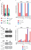
Figure 4. M-CSF Upregulates Host Anti-Apoptotic Genes to Mediate Resistance of Infected Macrophages to TRAIL
(A) mRNA levels for the anti-apoptotic genes Bfl-1 and Mcl-1 were measured by ribonuclease protection assay in HIV-1 wild type– and Δenv-infected macrophage cultures incubated in the presence or absence of M-CSF. (B) RNAi effectively mediated substantial reduction in Bfl-1 and Mcl-1 protein levels in human macrophages. Protein levels were determined by Western blotting and normalized to signals from tubulin. (C) RNAi-mediated Bf1–1 and Mcl-1 knockdown of mRNAs. mRNA levels, determined by quantitative RT-PCR, are shown as percentage relative to a scrambled siRNA (SCR1) (error bars, SD). (D) Bfl-1 and Mcl-1 are required for protection from TRAIL-mediated apoptosis. Macrophages were transfected with Mcl-1, Bfl-1, or a nontargeting control (SCR1) siRNA. Sixteen hours later, macrophages were treated with soluble TRAIL, or an antibody to M-CSF, and apoptosis was determined by ELISA for active (cleaved) caspase 3 (error bars, SD).
Figure 5
Figure 5. Imatinib Blocks M-CSF Signaling and Restores TRAIL Sensitivity of Infected Macrophages
(A) Imatinib inhibits M-CSF–dependent autophosphorylation of the M-CSF receptor. Macrophages were incubated for 16 h with 2 μM Imatinib mesylate before stimulation with recombinant M-CSF for 5 min. The M-CSF receptor was immunoprecipitated from cell lysates, and M-CSF–dependent tyrosine autophosphorylation was determined by Western blotting and densitometry. (B) Imatinib upregulates TRAIL-R1 expression on wild-type–infected macrophages. HIV-1 wild-type– and Δenv-infected macrophages were incubated with Imatinib for 16 h and TRAIL-R1 levels were determined by flow cytometry. (C) Macrophages from three donors were treated as in (B), and statistical analysis was performed on the mean expression of TRAIL-R1 in response to Imatinib (ANOVA; error bars, SEM). (D) Imatinib restores sensitivity to TRAIL. Macrophages were incubated with Imatinib for 16 h, challenged with soluble TRAIL, or both. Apoptotic HIV-1–infected and uninfected cells in the same cultures were determined 6 h later by Annexin V and propidium iodide staining and flow cytometry. Apoptotic cells were Annexin V+, propidium iodide (lower right quadrant). Imatinib renders HIV-1 wild-type–infected macrophages susceptible to TRAIL-mediated apoptosis. (E) Macrophages were treated as in (D), except apoptosis was determined 16 h after TRAIL exposure by ELISA for apoptotic histone-associated cellular DNA fragmentation in macrophage lysates (cell death ELISA; error bar, SD).
Figure 6
Figure 6. Prolonged Exposure to Imatinib Alone Is Sufficient to Induce Apoptosis in HIV-1–Infected Macrophages
Five days after infection with HIV-1LAI wild-type or Δenv viruses, macrophages were incubated with Imatinib for 24 or 120 h, and levels of apoptosis were determined by ELISA for histone-associated cellular DNA fragmentation in macrophage lysates (cell death ELISA; error bars, SD).
Figure 7
Figure 7. Experimental Model: Following HIV-1 Infection, De Novo Synthesized Viral Envelope Induces the Release of M-CSF
M-CSF opposes TRAIL by suppressing TRAIL-R1 expression and upregulating the anti-apoptotic factors Bfl-1 and Mcl-1. The anti-cancer drug Imatinib disables the protective effect of envelope by inhibiting M-CSF activity. Upon inhibition of M-CSF activity, TRAIL-R1 expression is increased and the action of Bfl-1 and Mcl-1 is blocked by pro-apoptotic bcl-2 family proteins. Further exposure of infected macrophages to Imatinib for several days induces apoptosis in the absence of TRAIL and is associated with increased pro-apoptotic protein expression.

Similar articles

Cited by

References

    1. Ray N, Doms RW. HIV-1 coreceptors and their inhibitors. Curr Top Microbiol Immunol. 2006;303:97–120. - PubMed
    1. Cicala C, Arthos J, Ruiz M, Vaccarezza M, Rubbert A, et al. Induction of phosphorylation and intracellular association of CC chemokine receptor 5 and focal adhesion kinase in primary human CD4+ T cells by macrophage-tropic HIV envelope. J Immunol. 1999;163:420–426. - PubMed
    1. Cicala C, Arthos J, Selig SM, Dennis G, Hosack DA, et al. HIV envelope induces a cascade of cell signals in non-proliferating target cells that favor virus replication. Proc Natl Acad Sci U S A. 2002;99:9380–9385. - PMC - PubMed
    1. Kaplan MJ, Ray D, Mo RR, Yung RL, Richardson BC. TRAIL (Apo2 ligand) and TWEAK (Apo3 ligand) mediate CD4+ T cell killing of antigen-presenting macrophages. J Immunol. 2000;164:2897–2904. - PubMed
    1. Martoglio B, Graf R, Dobberstein B. Signal peptide fragments of preprolactin and HIV-1 p-gp160 interact with calmodulin. EMBO J. 1997;16:6636–6645. - PMC - PubMed

Publication types

MeSH terms