. 2007 Sep 7;3(9):1291-307.
doi: 10.1371/journal.ppat.0030136.

Members of a large retroposon family are determinants of post-transcriptional gene expression in Leishmania

Affiliations

Members of a large retroposon family are determinants of post-transcriptional gene expression in Leishmania

Frédéric Bringaud et al. PLoS Pathog. .

Abstract

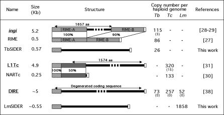
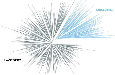
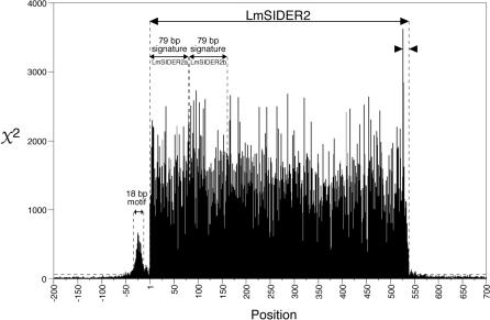
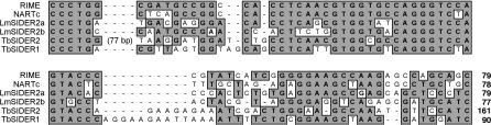
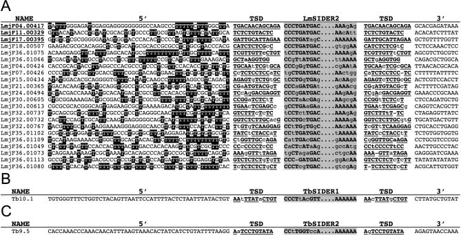
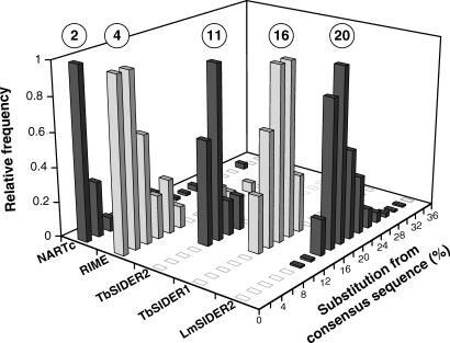
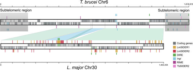
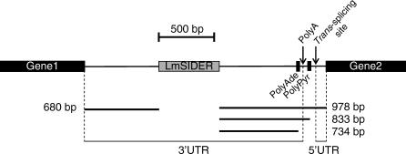
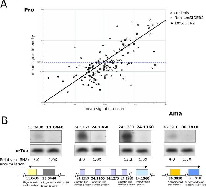
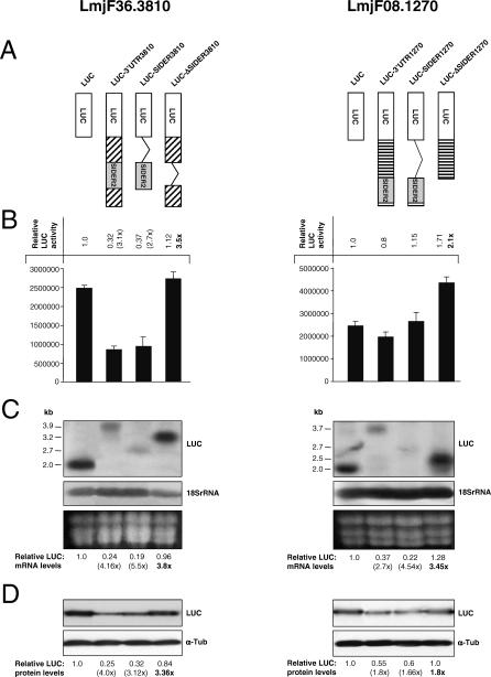
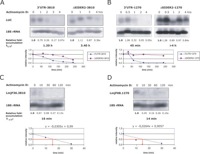
Trypanosomatids are unicellular protists that include the human pathogens Leishmania spp. (leishmaniasis), Trypanosoma brucei (sleeping sickness), and Trypanosoma cruzi (Chagas disease). Analysis of their recently completed genomes confirmed the presence of non-long-terminal repeat retrotransposons, also called retroposons. Using the 79-bp signature sequence common to all trypanosomatid retroposons as bait, we identified in the Leishmania major genome two new large families of small elements--LmSIDER1 (785 copies) and LmSIDER2 (1,073 copies)--that fulfill all the characteristics of extinct trypanosomatid retroposons. LmSIDERs are approximately 70 times more abundant in L. major compared to T. brucei and are found almost exclusively within the 3'-untranslated regions (3'UTRs) of L. major mRNAs. We provide experimental evidence that LmSIDER2 act as mRNA instability elements and that LmSIDER2-containing mRNAs are generally expressed at lower levels compared to the non-LmSIDER2 mRNAs. The considerable expansion of LmSIDERs within 3'UTRs in an organism lacking transcriptional control and their role in regulating mRNA stability indicate that Leishmania have probably recycled these short retroposons to globally modulate the expression of a number of genes. To our knowledge, this is the first example in eukaryotes of the domestication and expansion of a family of mobile elements that have evolved to fulfill a critical cellular function.


Conflict of interest statement

Competing interests. The authors have declared that no competing interests exist.

Figures

Figure 1
Figure 1. Description and Copy Number of the Trypanosomatid Retroposons
Retroelement names and sizes are indicated in the left margin. The names of coding or potentially coding retroposons (including the few retrotransposition-competent ingi and L1Tc elements) are underlined and boldfaced; the other elements are short non-autonomous retroposons (RIME, NARTc, TbSIDER, and LmSIDER). The central panel (“Structure”) represents the schematic map of the retroelements highlighting nucleotide sequence conservation, such as for the T. brucei ingi/RIME and T. cruzi L1Tc/NARTc pairs. The grey boxes represent conserved sequences between autonomous and non-autonomous members of a pair (the percentage of identity is indicated below), the hatched boxes represent the 79-bp sequence conserved at the 5′-extremity (“79 bp signature”), and the white boxes indicate the adenosine-rich stretch terminal sequences. The right panel indicates the number of each retroelement per haploid genome, including minichromosomes for T. brucei (dashes indicate the absence of elements in the corresponding genome). For autonomous elements, the value in brackets indicates the number (per haploid genome) of potentially functional elements, which may code for their own retrotransposition. Lm, L. major; Tb, T. brucei; Tc, T. cruzi.
Figure 2
Figure 2. Minimum Evolution Phylogenetic Tree of 785 LmSIDER Sequences
Only the LmSIDER sequences longer than 400 bp and smaller than 700 bp were considered to produce an alignment as described in Materials and Methods. The unrooted phylogenetic tree displays 140 LmSIDER1 (blue cluster) and 645 LmSIDER2 (black cluster) sequences.
Figure 3
Figure 3. The χ 2 Values for Individual Positions of the LmSIDER2 Core Sequence (538 bp) and Adjacent Regions (200 bp Upstream and 160 bp Downstream)
The χ 2 values were calculated as described in Materials and Methods from the set of 1,013 aligned LmSIDER2. The base composition of the whole L. major genome sequence was used to determine the background base distribution. The χ 2 values above the broken horizontal line correspond to significance levels of p < 0.001 for three degrees of freedom. Both “79 bp signatures,” called LmSIDER2a and LmSIDER2b, are positioned. The adenosine-rich stretch and the 18-bp thymidine-rich motif located at the 3′-extremity and upstream of the LmSIDER2, respectively, are indicated between arrowheads.
Figure 4
Figure 4. Comparison of the “79 bp Signature” Consensus Sequences between Different Trypanosomatid Retroposons
The first 79 bp of RIME, 78 bp of NARTc, 161 bp of TbSIDER2, 90 bp of TbSIDER1, and both “79 bp signatures” located at the 5′-extremity of the LmSIDER core sequence (LmSIDER2a and LmSIDER2b) were aligned, with the introduction of gaps (-) to maximize the alignments. Identical residues are shaded in grey.
Figure 5
Figure 5. Comparison of the Target Site Duplication (TSD) Flanking LmSIDER2 (A), TbSIDER1 (B), and TbSIDER2 (C) Sequences
In the left margin, the name of the element is indicated (the chromosome number is followed by the locus number). In this figure, only the LmSIDER/TbSIDER elements flanked by a TSD presenting two mismatches at the most are shown. The underlined names mean that the corresponding element is flanked by conserved TSD. The alignment of all the selected sequences was based on the retroelement sequences (grey column headed “LmSIDER2/TbSIDER1/TbSIDER2”) from which only the first 10 bp and the last 6 bp are shown (the conserved residues of the retroposon are boldfaced and capital characters). The TSD flanking the retroelements is indicated by boldfaced and underlined capital characters for the conserved residues. Lowercase characters in the TSD column correspond to nonconserved residues. In (A), T residues within the 5′-flanking sequences (called “5′”) that are abundant upstream of the TSD are indicated with white characters on a black background.
Figure 6
Figure 6. Divergence between Members of LmSIDER2 (1,013 Copies), TbSIDER1 (10 Copies), TbSIDER2 (12 Copies), RIME (70 Copies), and NARTc (115 Copies)
Bases covered by the L. major (LmSIDER2), T. cruzi (NARTc), and T. brucei (RIME, TbSIDER1, and TbSIDER2) short retroposons were sorted by their divergence from their consensus sequence. The consensus sequences, determined from the alignment of the core sequence of all the analyzed retroposons, approximate the element's original sequence at the time of insertion. The number of retroposons per fraction of 2% divergence is expressed as a fraction of the highest value, for which an arbitrary value of 1 has been assigned. The percentage of divergence was calculated using the matching region of the consensus sequences. The circled numbers on the top indicate the median value of each graph.
Figure 7
Figure 7. Comparative Analysis of TbChr6 and LmChr30 Syntenic Chromosomes
The syntenic regions between the L. major Chromosome 30 (LmChr30) and the T. brucei Chromosome 6 (TbChr6) are represented by blue diamonds. The grey shaded extremities of TbChr6 represent subtelomeric regions primarily composed of pseudogenes. The position of protein-coding genes and retroposons in each chromosome is indicated by vertical bars with the color code displayed on the right margin. Protein-encoding genes and ingi and DIRE retroposons are shown on the upper or lower part of the schematic chromosomes, depending on their strand location. Above or below the schematic chromosomes, the other retroposons (LmSIDER1, LmSIDER2, TbSIDER2, and RIME) are indicated, as are the blue and green arrows, which show the position of ingi and DIRE retroposons, respectively. The size (bp) of the chromosomes is indicated by the scale bars.
Figure 8
Figure 8. Predominant Localization of LmSIDERs in 3′UTRs
Within the intergenic region, trans-splicing generally occurs at an AG dinucleotide (trans-splicing site) downstream of a long polypyrimidine tract (PolyPyr). Polyadenylation (PolyA) of the upstream cistron takes place possibly as part of a coupled process together with trans-splicing. Consequently, the 3′UTR of the gene1 mRNA (upstream) ends at the putative polyadenylation site (PolyAde), and the 5′UTR of the gene2 mRNA (downstream) starts at the trans-splicing site. This figure shows the average relative position of the polyadenylation sites and the polypyrimidine tracts estimated with a previously developed algorithm [9], as well as the position of the LmSIDERs. The median size between LmSIDERs and the stop codon of the upstream gene (gene1), PolyAde, PolyPyr, or the start codon of the downstream gene (gene2) are also indicated.
Figure 9
Figure 9. LmSIDER2-Containing mRNAs Are Expressed for the Most Part at Lower Levels Relative to Transcripts Lacking LmSIDER2
(A) Log-scale scatter plot comparing ratios of normalized hybridization intensities between fluorescently labeled L. major promastigotes (Cy3) and amastigotes isolated from mice lesions (Cy5) RNA samples. A custom-designed low density DNA oligonucleotide-based microarray comprising 154 L. major genes, from which only 38 are predicted to harbor LmSIDER2 in their 3′UTR, was used for this study. Total RNA was purified from L. major promastigotes (Pro) grown to mid-log phase and from L. major lesion amastigotes (Ama). Probes were synthesized from total RNA and hybridized to microarrays in quadruplicate. Hybridization experiments were scanned and analyzed using recommended statistic parameters for low spot density arrays in the GeneSpring software. Mean signal intensities of all spots corresponding to one gene were background substracted and normalized with the mean spot intensity of the alien RNA NAC1. Genes were considered as differentially regulated when their expression ratios satisfied a p-value below 0.05. The mean signal intensity within the array was calculated to be ∼2,000 (horizontal dotted line). 50% of the genes were identified as significantly differentially expressed. 75% of the LmSIDER2-containing transcripts showed signal intensity lower than the mean intensity of all the spots as compared to 40% for the non-SIDER2 transcripts. (B) The steady-state levels of four pairs of transcripts that are part of the same transcription unit on three different L. major chromosomes were estimated by quantitative northern blotting. LmjF13.0440, LmjF24.1260, LmjF24.1360, and LmjF36.3810 transcripts (in bold) harbor LmSIDER2 in their 3′UTR, whereas LmjF13.0430, LmjF24.1250, LmjF24.1280, and LmjF36.3910 do not. LmjF13.0430 and LmjF13.0440 genes are tandemly linked on Chromosome 13, LmjF24.1250 and LmjF24.1260 are tandemly linked on Chromosome 24, and LmjF24.1280 and LmjF24.1360 genes (chr 24) and LmjF36.3810 and LmjF36.3910 genes (chr 36) are separated by seven and eight genes, respectively. The predicted putative function of the above protein-coding genes is indicated at the lower panel. Equal amounts (∼20 μg) of total RNA were loaded on agarose gel prior to transfer onto a nylon membrane and hybridized with gene-specific probes that were of the same length, GC content, and labeling activity. mRNA levels were quantitated with respect to the amount of total RNA loaded on the gel as verified by hybridization using the alpha-tubulin (α-Tub) gene-specific probe. The normalized numbers are indicated below the blots.
Figure 10
Figure 10. LmSIDER2 Promotes mRNA Downregulation in L. major
The potential role of LmSIDER2-containing LmjF36.3810 and LmjF08.1270 3′UTRs in regulating either mRNA or protein levels was evaluated in L. major recombinant parasites grown as promastigotes. (A) Schematic representation of the different LUC chimeric constructs used in this study with the corresponding name indicated on the top. The cross-hatched boxes indicate 3′UTR sequences other than the SIDER sequence. In all the LUC-expressing vectors, the LUC transcript is processed at the 5′-end by sequences from the alpha-tubulin intercistronic region that provides signals for trans-splicing (see Materials and Methods). A series of 3′UTR sequences comprising either the full-length 3′UTR (3′UTR-3810, 3′UTR-1270) from the LmjF36.3810 (3,810) and LmjF08.1270 (1,270) transcripts, or the defined LmSIDER2 sequence alone (SIDER-3810, SIDER-1270), or the 3′UTR lacking the LmSIDER2 element (ΔSIDER-3810, ΔSIDER-1270), were cloned downstream of the reporter gene firefly luciferase (LUC). The presence and location of LmSIDER2 within the 3′UTR of LmjF08.1270 and LmjF36.3810 transcripts was verified by 3′UTR mapping. The pSPYNEOαLUC vector (LUC) lacking any regulatory 3′UTR region [16] was used here as a control. The above LUC constructs were introduced by electroporation into the L. major LV39 strain to obtain stable recombinant parasites. The copy number of the different LUC vectors per cell was similar as estimated by Southern blot hybridization (unpublished data). (B) LUC activity was measured as indicated in Materials and Methods. The data were normalized relatively to the control transfectant (LUC). Numbers in parentheses correspond to fold differences in LUC activity with respect to the control LUC strain. The number in bold represents the fold difference in LUC activity compared to the full-length 3′UTR-3810 or 3′UTR-1270. Values are mean + standard error of four independent experiments. (C) Northern blot analysis of total RNA extracted from L. major promastigotes expressing the different LUC-chimeric constructs described in (A). RNA loading on the gel was monitored by hybridization to the 18S rRNA–specific probe. A section of the ethidium-stained gel containing the three ribosomal RNAs is also shown to demonstrate loading. Northern blot hybridization was repeated at least two times and similar results were obtained. (D) Western blot analysis of total protein lysates from L. major–LUC recombinant promastigotes using the anti-LUC antibody. Membranes were stripped and reacted with an anti-alpha-tubulin (α-Tub) antibody to verify protein loading. Western blot analyses were carried out with three different cultures for each transfectant and similar results were obtained. Numbers in parentheses correspond to fold differences relative to the LUC control protein steady-state levels.
Figure 11
Figure 11. LmSIDER2 Is Involved in mRNA Destabilization
(A and B) Comparison of mRNA half-lives (t1/2) in L. major promastigotes between LmSIDER2-containing LUC chimeric mRNAs, 3′UTR-3810 (A) and 3′UTR-1270 (B), and LUC mRNAs lacking the LmSIDER2 element, ΔSIDER2–3810 (A) and ΔSIDER2–1270 (B). Approximately 107 L. major recombinant promastigotes/ml were treated with 10 μg/ml of actinomycin D, an inhibitor of de novo transcription, and RNA was isolated at the time points shown and analyzed by northern blotting. Transcript levels were normalized with respect to the amount of the rRNA loaded using the 18S rRNA probe as an internal control. The levels of mRNAs were assessed using phosphorimaging. The values shown below the blots represent the LUC mRNA fold accumulation with respect to its abundance prior to the addition of actinomycin D (0). This is a representative experiment out of two that showed very similar results. (C and D) The decay of the endogenous LmjF36.3810 (C) and LmjF08.1270 (D) transcripts was also assessed in L. major promastigotes using actinomycin D (using northern blotting). The half-lives of LmSIDER2-containing LUC chimeric mRNAs and LmjF36.3810 and LmjF08.1270 endogenous transcripts were estimated based on hybridization intensities with gene-specific probes normalized with the 18S rRNA probe with respect to different time points of actinomycin D treatment. Experiments shown here are representative of two that showed very similar results.

Similar articles

Cited by

References

    1. Haag J, O'Huigin C, Overath P. The molecular phylogeny of trypanosomes: Evidence for an early divergence of the Salivaria. Mol Biochem Parasitol. 1998;91:37–49. - PubMed
    1. Berriman M, Ghedin E, Hertz-Fowler C, Blandin G, Renauld H, et al. The genome of the African trypanosome Trypanosoma brucei. Science. 2005;309:416–422. - PubMed
    1. El-Sayed NM, Myler PJ, Bartholomeu DC, Nilsson D, Aggarwal G, et al. The genome sequence of Trypanosoma cruzi, etiologic agent of Chagas disease. Science. 2005;309:409–415. - PubMed
    1. Ivens AC, Peacock CS, Worthey EA, Murphy L, Aggarwal G, et al. The genome of the kinetoplastid parasite, Leishmania major. Science. 2005;309:436–442. - PMC - PubMed
    1. Liang XH, Haritan A, Uliel S, Michaeli S. trans and cis splicing in trypanosomatids: Mechanism, factors, and regulation. Eukaryot Cell. 2003;2:830–840. - PMC - PubMed

Publication types