. 2007 Sep 7;3(9):1308-19.
doi: 10.1371/journal.ppat.0030138.

Structure of the malaria antigen AMA1 in complex with a growth-inhibitory antibody

Affiliations

Structure of the malaria antigen AMA1 in complex with a growth-inhibitory antibody

Andrew M Coley et al. PLoS Pathog. .

Erratum in

  • PLoS Pathog. 2007 Nov;3(11):e172

Abstract

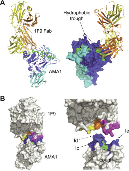
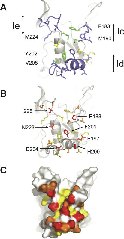
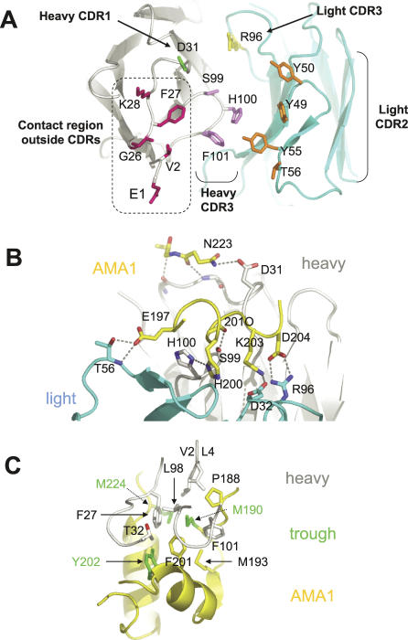
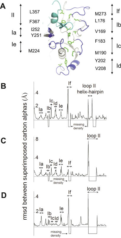
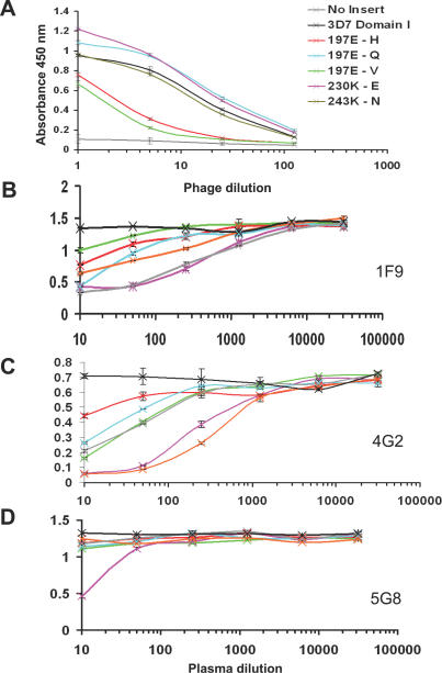
Identifying functionally critical regions of the malaria antigen AMA1 (apical membrane antigen 1) is necessary to understand the significance of the polymorphisms within this antigen for vaccine development. The crystal structure of AMA1 in complex with the Fab fragment of inhibitory monoclonal antibody 1F9 reveals that 1F9 binds to the AMA1 solvent-exposed hydrophobic trough, confirming its importance. 1F9 uses the heavy and light chain complementarity-determining regions (CDRs) to wrap around the polymorphic loops adjacent to the trough, but uses a ridge of framework residues to bind to the hydrophobic trough. The resulting 1F9-AMA1-combined buried surface of 2,470 A(2) is considerably larger than previously reported Fab-antigen interfaces. Mutations of polymorphic AMA1 residues within the 1F9 epitope disrupt 1F9 binding and dramatically reduce the binding of affinity-purified human antibodies. Moreover, 1F9 binding to AMA1 is competed by naturally acquired human antibodies, confirming that the 1F9 epitope is a frequent target of immunological attack.


Conflict of interest statement

Competing interests. The authors have declared that no competing interests exist.

Figures

Figure 1
Figure 1. Structure of AMA1 in Complex with 1F9 Fab
(A) Alternate views of AMA1 in complex with 1F9 Fab. Backbone trace of 1F9 Fab with the heavy chain coloured orange and the light chain coloured yellow. Backbone trace of AMA1 (left) or surface view (right) with domain I coloured dark blue and domain II coloured light blue. The AMA1 hydrophobic trough is coloured green. This figure and other figures depicting the structure were generated using PyMOL [56]. (B) Surface representation of the 1F9 Fab bound to AMA1. View of the complex (left), or 1F9 and AMA1 separated (right). Light chain residues that contact AMA1 (within 4 Å) are coloured yellow. Heavy chain residues that contact AMA1 are coloured orange (residues from the CDR loops) or pink (framework residues). AMA1 residues within 4 Å of 1F9 are coloured dark blue, with the hydrophobic trough residues contacting 1F9 coloured green. AMA1 loops Ic, Id, and Ie are indicated.
Figure 2
Figure 2. 1F9 Epitope on AMA1
(A,B) Show identical views of the epitope with side chains that interact with AMA1 with a buried surface area of 7 Å2 or more shown in stick form. (C) Is the same view but showing the surface of the epitope. (A) Hydrophobic trough residues that contact 1F9 are shown in green with their residue numbers on either side of the figure. Loops Ic, Id, and Ie and their 1F9-contacting residues are shown in blue. Side chains that extend from the PAN domain scaffold and interact with 1F9 are coloured yellow. (B,C) Principal contact residues. Side chains contacting 1F9 with a surface area of greater than 78 Å2 or more are coloured red. Side chains with a contact surface area of between 68 and 38 Å2 are coloured orange. Side chains with a contact surface area of between 24 and 7 Å2 are coloured yellow. Surface area interactions were calculated using AREAIMOL [54].
Figure 3
Figure 3. Effect of AMA1 Mutations on 1F9 Binding
(A) Mapping of point and deletion mutations onto the AMA1 structure. Point mutations at positions 197, 200, 201, 204, and 225 (coloured red) disrupted 1F9 binding. Point mutations at position 228 (yellow) were partially disruptive. Point mutations at positions 196, 230, 243, and 244 (blue) had no effect on 1F9 binding. N-terminal deletion of loop Ic (blue) had no effect on binding [38]. (B) M13 phage expressing point mutations in domain I AMA1 were added to immobilized 1F9 at a series of dilutions. Bound phage were assayed by the addition to peroxidase-conjugated anti-M13 mAb followed by a colourimetric assay. Assays were carried out in duplicate and the error bars indicate the two measured absorbance values.
Figure 4
Figure 4. 1F9 Recognition Surface and Details of the 1F9–AMA1 Interaction
In all panels AMA1 main chain is coloured yellow, 1F9 light chain is light blue, and 1F9 heavy chain is grey/white. (A) View of the CDR loops on 1F9. Residues shown are those with an AMA1 contact surface area of at least 35 Å2. Light chain CDR2 and 3 side chains are coloured orange and yellow, respectively, with heavy chain CDR 1 and 3 side chains coloured green and violet, repectively. Residues coloured pink are in the antibody framework region. Amino acid numbering refers to the 1F9 sequence. In the Kabat numbering scheme [57] heavy chain residue S99 is S95, H100 is H96, and F101 is F102. Light chain numbering is the same as in the Kabat numbering scheme. (B) Principal hydrogen bond interactions between AMA1 and 1F9. Side view of the AMA1-1F9 interface showing that most hydrogen bond interactions occur between AMA1 loop Id (residues 197–204), 1F9 light chain residues and the heavy chain CDR3 (residues S99 and H100). 201O is the main-chain carbonyl oxygen of residue 201. (C) View of the 1F9 heavy chain hydrophobic residues sitting in the hydrophobic trough. Hydrophobic trough side chains are coloured green.
Figure 5
Figure 5. Comparison of Free and 1F9-Complexed AMA1 Structures
(A) Structure of the hydrophobic trough and surrounding loops. Domain I loops, Ia-If, are shown in dark blue. The domain II loop, II, is shown in light blue. The 12 hydrophobic trough residues are shown in green with the label on either side of the figure showing the amino acid and numbering. I252 and Y251 are not adjacent to loop Ia, but other trough residue labels are shown next to the loop they are associated with. (B–D) Comparison of the main-chain trajectories of AMA1 structures.rmsds between superimposed carbon alphas were calculated using the program LSQKAB [54] and plotted for each residue. (A) Comparison between AMA1 model 1Z40 and AMA1-1F9 crystal form 1. (B) Comparison between AMA1 model 1Z40 and AMA1-1F9 crystal form 2. (C) Comparison between AMA1-1F9 crystal forms 1 and 2. In instances where residues were missing in either of the compared models the rmsd was assigned a value of −1.
Figure 6
Figure 6. Binding of Human Plasma to AMA1
(A) Point mutations abrogate binding of AMA1 domain I to human antibodies. Phage expressing AMA1 domain I were tested for their ability to bind to immobilised AMA1-affinty purified human antibodies from a pool of Papua New Guinean blood donors. Control (black) is M13 phage expressing strain 3D7 AMA1 domain I. Negative control (grey) is phage expressing no insert. Point mutations tested were: residue 197E changed to (H red, Q cyan, V green), or residue 230 K-E (violet), or residue 243 K-N. Assays were carried out in duplicate and the error bars indicate the two measured absorbance values. (B–D) Individual human plasma compete for 1F9 binding to AMA1. Full-length 3D7 AMA1 ectodomain was immobilised on plastic. MAbs 1F9 (A), 4G2 (B), and 5G8 (C) were allowed to bind and the ability of a set of human plasma to compete for mAb binding was tested: plasma P8 (green), plasma P45 (pink), plasma P60 (red), plasma P69 (light blue), plasma P111 (orange), and plasma M157 (grey). Pooled plasma derived from a malaria unexposed individuals served as a negative control (black). Assays were carried out in duplicate and error bars indicate the two measured absorbance values.

Similar articles

Cited by

References

    1. Breman JG, Alilio MS, Mills A. Conquering the intolerable burden of malaria: What's new, what's needed: a summary. Am J Trop Med Hyg. 2004;71:1–15. - PubMed
    1. Sachs J, Malaney P. The economic and social burden of malaria. Nature. 2002;415:680–685. - PubMed
    1. Baird JK. Host age as a determinant of naturally acquired immunity to Plasmodium falciparum. Parasitol Today. 1995;11:105–111. - PubMed
    1. Cohen S, McGregor IA, Carrington SC. Gamma-globulin and acquired immunity to human malaria. Nature. 1961;192:733–737. - PubMed
    1. Cohen S, Butcher GA. Properties of protective malarial antibody. Nature. 1970;225:732–734. - PubMed

Publication types