

Unsupervised border detection in dermoscopy images
- PMID: 17908199
- PMCID: PMC3191533
- DOI: 10.1111/j.1600-0846.2007.00251.x
Unsupervised border detection in dermoscopy images
Abstract
Background: As a result of the advances in skin imaging technology and the development of suitable image processing techniques, during the last decade, there has been a significant increase of interest in the computer-aided diagnosis of skin cancer. Automated border detection is one of the most important steps in this procedure as the accuracy of the subsequent steps crucially depends on the accuracy of this step.
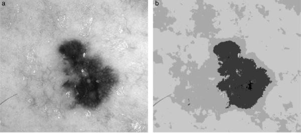
Methods: In this article, we present an unsupervised approach to border detection in dermoscopy skin lesion images based on a modified version of the JSEG algorithm.
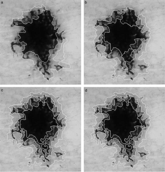
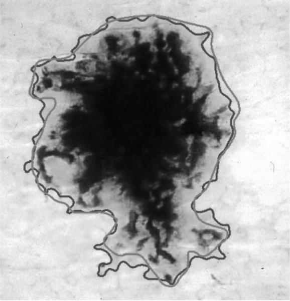
Results: The method is tested on a set of 100 dermoscopy images. The border detection error is quantified by a metric that uses manually determined borders from a dermatologist as the ground truth. The results are compared with three other automated methods and manually determined borders by a second dermatologist.
Conclusion: The results demonstrate that the presented method achieves both fast and accurate border detection in dermoscopy images.
Figures
References
-
- Jemal A, Murray T, Ward E, et al. Cancer statistics 2005. CA: Cancer J Clinicians. 2005;55:10–30. - PubMed
-
- Mayer J. Systematic review of the diagnostic accuracy of dermoscopy in detecting malignant melanoma. Med J Aust. 1997;167:206–210. - PubMed
-
- Kittler H, Pehamberger K, Wolff K, Binder M. Diagnostic accuracy of dermoscopy. Lancet Oncol. 2002;3:159–165. - PubMed
-
- Stolz W, Braun-Falco O, Bilek P, Landthaler M, Burgdorf WHC, Cognetta AB. Color atlas of dermatoscopy. 2nd edn. Blackwell Wissenschafts-Verlag; Berlin, Germany: 2002.
-
- Binder M, Schwarz M, Winkler A, Steiner A, Kaider A, Wolff K, Pehamberger H. Epiluminescence microscopy. A useful tool for the diagnosis of pigmented skin lesions for formally trained dermatologists. Arch Dermatol. 1995;131:286–291. - PubMed
Publication types
MeSH terms
Grants and funding
LinkOut - more resources
Full Text Sources
Other Literature Sources
Medical
Research Materials
