. 2007 Dec;103(6):2268-80.
doi: 10.1111/j.1471-4159.2007.04931.x. Epub 2007 Oct 1.

The regulation of M1 muscarinic acetylcholine receptor desensitization by synaptic activity in cultured hippocampal neurons

Affiliations

The regulation of M1 muscarinic acetylcholine receptor desensitization by synaptic activity in cultured hippocampal neurons

Jonathon M Willets et al. J Neurochem. 2007 Dec.

Abstract

To better understand metabotropic/ionotropic integration in neurons we have examined the regulation of M1 muscarinic acetylcholine (mACh) receptor signalling in mature (> 14 days in vitro), synaptically-active hippocampal neurons in culture. Using a protocol where neurons are exposed to an EC(50) concentration of the muscarinic agonist methacholine (MCh) prior to (R1), and following (R2) a desensitizing pulse of a high concentration of this agonist, we have found that the reduction in M(1) mACh receptor responsiveness is decreased in quiescent (+tetrodotoxin) neurons and increased when synaptic activity is enhanced by blocking GABA(A) receptors with picrotoxin. The picrotoxin-mediated effect on M1 mACh receptor responsiveness was completely prevented by alpha-amino-3-hydroxy-5-methyl-4-isoxazolepropionic acid receptor blockade. Inhibition of endogenous G protein-coupled receptor kinase 2 by transfection with the non-G(q/11)alpha-binding, catalytically-inactive (D110A,K220R)G protein-coupled receptor kinase 2 mutant, decreased the extent of M1 mACh receptor desensitization under all conditions. Pharmacological inhibition of protein kinase C (PKC) activity, or chronic phorbol ester-induced PKC down-regulation had no effect on agonist-mediated receptor desensitization in quiescent or spontaneously synaptically active neurons, but significantly decreased the extent of receptor desensitization in picrotoxin-treated neurons. MCh stimulated the translocation of diacylglycerol- sensitive eGFP-PKCepsilon, but not Ca2+/diacylglycerol-sensitive eGFP-PKCbetaII in both the absence, and presence of tetrodotoxin. Under these conditions, MCh-stimulated eGFP-myristoylated, alanine-rich C kinase substrate translocation was dependent on PKC activity, but not Ca2+/calmodulin. In contrast, picrotoxin-driven translocation of myristoylated, alanine-rich C kinase substrate was accompanied by translocation of PKCbetaII, but not PKCepsilon, and was dependent on PKC and Ca2+/calmodulin. Taken together these data suggest that the level of synaptic activity may determine the different kinases recruited to regulate M1 mACh receptor desensitization in neurons.


Figures

Fig. 1
Fig. 1
Effects of synaptic activity on M1 muscarinic acetylcholine (mACh) receptor responsiveness and re-sensitization assessed through inositol 1,4,5-trisphosphate (IP3) imaging of hippocampal neurons. The desensitization protocol (R1/Rmax/R2) was performed as described in the Experimental Procedures section. Representative traces showing M1 mACh receptor desensitization in the presence of (a) tetrodotoxin (TTx) (500 nmol/L); (b) no pre-addition (spontaneous synaptic activity; Spont in panel d); and (c) synaptic activity induced by picrotoxin (PiTx, 100 μmol/L). Picrotoxin was present 3 min prior to and throughout the experiment. Methacholine (R1, 10 μmol/L, 30 s; Rmax, 100 μmol/L, 60 s; R2 10 μmol/L, 30 s) was added as indicated by the bars. (d) Cumulative data for time-courses of M1 mACh receptor re-sensitization in the absence (Spont) or presence of TTx, or following PiTx addition. Data are expressed as means ± SEM for the percentage change in R2 relative to the R1 response, for 5–15 neurons taken from at least three separate hippocampal preparations. Significant differences in the R2/R1 ratio from the +TTx condition at a given time-point are indicated as *p< 0.05; **p< 0.01.
Fig. 2
Fig. 2
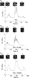
Effect of inhibition of α-amino-3-hydroxy-5-methyl-4-isoxazolepropionic acid and NMDA receptors on M1 muscarinic acetylcholine receptor desensitization. Representative images and traces showing M1 muscarinic acetylcholine receptor desensitization in the presence of (a) picrotoxin (PiTx, 100 μmol/L); (b) PiTx (100 μmol/L) + D-AP5 (50 μmol/L); (c) PiTx (100 μmol/L) + DNQX (10 μmol/L). Picrotoxin ± D-AP5/DNQX were present 3 min prior to R1 methacholine addition and throughout the experiment. methacholine (R1, 10 μmol/L, 30 s; Rmax, 100 μmol/L, 60 s; R2 10 μmol/L, 30 s) was added as indicated by the bars. Data are representative of 8–17 neurons taken from at least three separate hippocampal preparations (see Table 1).
Fig. 3
Fig. 3
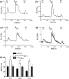
Effects of inhibiting endogenous G protein-coupled receptor kinase (GRK) 2 and GRK6 activities on M1 muscarinic acetylcholine (mACh) receptor responsiveness. Neurons were co-transfected with PH domain of PLCδ tagged to enhanced green fluorescent protein and empty vector (pcDNA3), D110A,K220RGRK2, or K215RGRK6. A standard R1/Rmax/R2 protocol was applied to assess M1 mACh receptor desensitization in either the absence (spontaneous activity; spont) or presence of tetrodotoxin (TTx, 500 nmol/L), or in the presence of picrotoxin (PiTx, 100 μmol/L). Representative traces showing M1 mACh receptor desensitization in the presence of (a) pcDNA3 and TTx; (b) D110A,K220RGRK2 and TTx; (c) pcDNA3 and PiTx; and (d) D110A,K220RGRK2 and PiTx. Methacholine addition is indicated by the horizontal bars. (e) cumulative data are presented as means ± SEM for the percentage change in R2 relative to the R1 response, for five neurons for each treatment taken from at least three separate hippocampal preparations. Significant differences in the R2/R1 ratio caused by expression of the dominant-negative D110A,K220RGRK2 or K215RGRK6 constructs are indicated as *p< 0.05; **p< 0.01.
Fig. 4
Fig. 4
Effects of manipulating protein kinase C activity in hippocampal neurons on M1 muscarinic acetylcholine receptor responsiveness. Neurons were co-transfected with PH domain of PLCδ tagged to enhanced green fluorescent protein and empty vector (pcDNA3) or D110A,K220RG protein-coupled receptor kinase (GRK) 2 and changes in receptor responsiveness determined using the standard R1/Rmax/R2 protocol (see Experimental Procedures). Neurons were pre-incubated with either vehicle (Control) or staurosporine (1 μmol/L; Stauro) for 15 min prior to, and throughout the experiment. Representative traces are shown for neurons treated with picrotoxin (PiTx, 100 μmol/L) in the presence of (a) pcDNA3 and vehicle; (b) D110A,K220RGRK2 and vehicle; (c) pcDNA3 + staurosporine (1 μmol/L); (d) D110A,K220RGRK2 and staurosporine (1 μmol/L). (e) cumulative data are shown for either spontaneously active or picrotoxin-treated neurons and are expressed as means ± SEM for the percentage change in R2 relative to the R1 response, for ≥ 5 neurons per treatment taken from at least three separate hippocampal preparations. Significant differences in the R2/R1 ratio caused by either D110A,K220RGRK2 expression are indicated as *p< 0.05; **p< 0.01, while a significant effect of staurosporine pre-treatment is shown as +p< 0.05.
Fig. 5
Fig. 5
Effects of Ca2+/calmodulin antagonism or protein kinase C inhibition on M1 muscarinic acetylcholine receptor-driven myristoylated, alanine-rich C kinase substrate (MARCKS) translocation in hippocampal neurons. Neurons were transfected with enhanced green fluorescent protein (eGFP)-MARCKS. Agonist addition [methacholine (MCh), 3 μmol/L] for 30 s induced a robust translocation of eGFP-MARCKS from the plasma membrane to the cytoplasm, which completely reversed after agonist removal. Following the initial MCh stimulation (S1), neurons were treated with W5 (25 μmol/L; panel a), W7 (25 μmol/L; panel b), or staurosporine (1 μmol/L; panel c) for 15 min, prior to a second application of MCh (3 μmol/L, for 30 s; S2). Panels (a)–(c) present representative traces with accompanying images. (d) cumulative data showing the effect of W5, W7 and staurosporine (Stauro) on the percentage change in S2 relative to the S1 for MCh-stimulated eGFP-MARCKS translocation. Data are expressed as means ± SEM for at least four neurons per treatment taken from three separate hippocampal preparations. The statistically significant effect of staurosporine treatment on the S2/S1 ratio is indicated as ***p< 0.001.
Fig. 6
Fig. 6
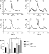
Effects of Ca2+/calmodulin antagonism and/or protein kinase C inhibition on picrotoxin (PiTx)-induced myristoylated, alanine-rich C kinase substrate translocation. Neurons were transfected with enhanced green fluorescent protein-myristoylated, alanine-rich C kinase substrate. Picrotoxin (100 μmol/L) was initially added to neurons for 2 min (S1); following washout neurons were incubated with vehicle (a), W5 (25 μmol/L; b), W7 (25 μmol/L; c), staurosporine (1 μmol/L; d) or W7 and staurosporine together (e), for 15 min, followed immediately by a second challenge (S2) with picrotoxin (100 μmol/L) for 2 min. Panels (a)–(e) present representative traces. (f) cumulative data are expressed as means ± SEM for the percentage change in S2 relative to the S1 response, for 4–6 neurons for each condition taken from at least three separate hippocampal preparations. Statistically significant inhibitor effects on the S2/S1 ratio compared to vehicle control are indicated as *p< 0.05; **p< 0.01.
Fig. 7
Fig. 7
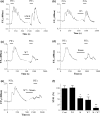
Translocation of conventional and novel protein kinase C (PKCs) following picrotoxin (PiTx) treatment of hippocampal neuron cultures. eGFP-PKCβII or eGFP-PKCε transfected neurons were challenged with PiTx (100 μmol/L) and PKC translocation to the plasma membrane determined as the decrease in cytosolic fluorescence. In panels (a) and (b), neurons were also loaded with the Ca2+-sensitive dye Fura-Red for 60 min prior to addition of PiTx. Note that downward deflections in the Ca2+ trace indicate increases in [Ca2+]i. (a) representative trace showing that picrotoxin addition alone did not stimulate translocation of eGFP-PKCε. (b) representative trace showing that, in contrast, PiTx addition alone caused transient increases in intracellular [Ca2+] that are mirrored by rapid and transient translocations of eGFP-PKCβII. (c) representative trace showing the effects of sequential methacholine (MCh, 100 μmol/L) and PiTx (100 μmol/L) additions on the translocation of eGFP-PKCβII. (d) representative trace showing the effects of the α-amino-3-hydroxy-5-methyl-4-isoxazolepropionic acid receptor blocker 6,7-dinitroquinoxaline-2,3-dione (DNQX, 10 μmol/L) and tetrodotoxin (TTx, 500 nmol/L) on PiTx-induced eGFP-PKCβII translocations. All representative traces are shown for experiments repeated on 4–10 coverslips from at least three separate hippocampal preparations.
Fig. 8
Fig. 8
Effects of synaptic activity on M1 muscarinic acetylcholine receptor responsiveness assessed at the level of protein kinase C (PKC) isoenzyme recruitment to the plasma membrane in hippocampal neurons. Neurons were transfected with eGFP-PKCε or eGFP-PKCβII and subjected to the standard R1/Rmax/R2 protocol in the absence (a), or presence (b and c) of picrotoxin (PiTx, 100 μmol/L) for 3 min prior to, and for the duration of the experiment. Methacholine (R1, 10 μmol/L, 30 s; Rmax, 100 μmol/L, 60 s; R2 10 μmol/L, 30 s) was added as indicated by the bars. (a) representative trace showing the eGFP-PKCε translocations evoked during the methacholine stimulation protocol. (b) representative traces showing the effects of PiTx (100 μmol/L) on plasma membrane recruitment of eGFP-PKCε and concurrent changes in intracellular [Ca2+], during the receptor desensitization protocol. Note that downward deflections in the Ca2+ trace reflect increases in [Ca2+]i. (c) representative traces showing the coincident translocation in eGFP-PKCβII to the plasma membrane with changes in intracellular [Ca2+]. All representative traces are shown for experiments repeated on 5–8 coverslips from at least three separate hippocampal preparations. (d) cumulative data showing the effects of spontaneous activity and picrotoxin (100 μmol/L)-stimulated synaptic activity on M1 muscarinic acetylcholine receptor desensitization measured using the standard R1/Rmax/R2 protocol. Data are presented as means ± SEM for the % change in R2 relative to the R1 response, for 6–18 neurons for each treatment taken from at least three separate hippocampal preparations. Statistically significant effects of PiTx addition on the R2/R1 ratio are indicated as **p< 0.01 (Student’s t-test).

Similar articles

Cited by

References

    1. Arbuzova A, Schmitz A A, Vergeres G. Cross-talk unfolded: MARCKS proteins. Biochem. J. 2002;362:1–12. - PMC - PubMed
    1. Bacci A, Verderio C, Pravettoni E, Matteoli M. Synaptic and intrinsic mechanisms shape synchronous oscillations in hippocampal neurons in culture. Eur. J. Neurosci. 1999;11:389–397. - PubMed
    1. Bartlett P J, Young K W, Nahorski S R, Challiss R A J. Single cell analysis and temporal profiling of agonist-mediated inositol 1,4,5-trisphosphate, Ca2+, diacylglycerol, and protein kinase C signaling using fluorescent biosensors. J. Biol. Chem. 2005;280:21837–21846. - PubMed
    1. Berkeley J L, Levey A I. Cell-specific extracellular signal-regulated kinase activation by multiple G protein-coupled receptor families in hippocampus. Mol. Pharmacol. 2003;63:128–135. - PubMed
    1. Berkeley J L, Gomeza J, Wess J, Hamilton S E, Nathanson N M, Levey A I. M1 muscarinic acetylcholine receptors activate extracellular signal-regulated kinase in CA1 pyramidal neurons in mouse hippocampal slices. Mol. Cell. Neurosci. 2001;18:512–524. - PubMed

Publication types

MeSH terms

LinkOut - more resources