. 2007 Oct 1:7:184.
doi: 10.1186/1471-2407-7-184.

Paradoxical antiproliferative effect by a murine mammary tumor-derived epithelial cell line

Affiliations

Paradoxical antiproliferative effect by a murine mammary tumor-derived epithelial cell line

Esteban N Gurzov et al. BMC Cancer. .

Abstract

Background: Despite significant advancement in breast cancer therapy, there is a great need for a better understanding of the mechanisms involved in breast carcinogenesis and progression, as well as of the role of epigenetic contributions from stromal cells in mammary tumorigenesis. In this study, we isolated and characterized murine mammary tumor-derived epithelial and myofibroblast cell lines, and investigated the in vitro and in vivo effect of cellular soluble factors produced by the epithelial cell line on tumor cells.

Methods: Morphology, immunophenotype, cytogenetics, invasiveness, and tumorigenicity of epithelial (LM-234ep) and myofibroblast (LM-234mf) cell lines isolated from two murine mammary adenocarcinomas with common ancestor were studied. The in vitro effects of LM-234ep conditioned medium on proliferation, cell cycle distribution, and expression of cell cycle proteins, were investigated in LM-234mf cells, mouse melanoma cells (B16-F10), and human cervical adenocarcinoma cells (HeLa). The in vivo anti-tumor activity of LM-234ep conditioned media was evaluated in subcutaneous tumors formed in nude mice by B16-F10 and HeLa cells.

Results: LM-234ep cells were found to be cytokeratin positive and hipertriploid, whereas LM-234mf cells were alpha-smooth muscle actin positive and hypohexaploid. Chromosome aberrations were found in both cases. Only LM-234mf revealed to be invasive in vitro and to secrete active MMP-2, though neither of the cell types were able to produce progressing tumors. LM-234ep-derived factors were able to inhibit the in vitro growth of LM-234mf, B16-F10, and HeLa cells, inducing cell cycle arrest in G0/G1 phase. The administration of LM-234ep conditioned medium inhibited the growth of B16-F10 and HeLa tumors in nude mice.

Conclusion: Our data suggest the existence of epithelial cell variants with tumor suppressive properties within mammary tumors. To our knowledge, this is the first report showing antiproliferative and antineoplastic activities induced by tumor-derived epithelial cells.


Figures
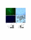
Figure 1
Figure 1
Alpha smooth muscle actin (SMA) and cytokeratin expression. SMA (green staining) was detected in LM-234mf (A) but not in LM-234ep (C) cells, using a FITC-conjugated primary antibody. Nuclei (blue staining) were identified using DAPI. Cytokeratin was undetectable in LM-234mf (B), whereas strongly expressed in LM-234ep (D), as revealed by immunohistochemistry using an anti-Pan cytokeratin antibody. Magnification bar, 100 μm. Cell lysates (25 μg/lane) were analyzed using Western blot for cytokeratins (E) and αSMA (F). The cytokeratins detected with the anti-pan antibody used range between 40 and 68 kDa. The band immunodetected for αSMA has an approximate molecular weight of 42 kDa. Glyceraldehyde 3-Phosphate Dehydrogenase (GAPDH) was used as a loading control.
Figure 2
Figure 2
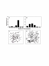
Chromosome analysis of LM-234 cell lines. Histograms showing the chromosome number distribution in LM-234ep (A) and LM-234mf (B) cells. A minimum of 50 metaphase spreads was analyzed in each case. Representative metaphases of LM-234ep (C) and LM-234mf (D) cells. Arrows point different chromosomal aberrations including: acentric fragments (1), chromosome break (2), and centromeric fusion (3). Magnification, × 1000.
Figure 3
Figure 3
Gelatinase expression/activity and invasive abilities of LM-234 cell lines. Conditioned media derived from LM-234mf and LM-234ep cells were analyzed by gelatin zymography, using 7F2 and BMA3.1A7 cell lines as positive controls for mouse MMP-2 and MMP-9, respectively (A). LM-234mf and ep cells were compared for their ability to invade through Matrigel-coated Transwell 8 μm-pore filters (B). The number of cells traversing the membrane was quantified and expressed as mean ± SE (n = 3).
Figure 4
Figure 4
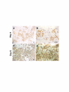
LM-234ep cells overgrow LM-234mf cells. Cells were co-cultured at a 1:1 ratio and immunostained for cytokeratin on days 4 (A and B) and 8 (C and D) to distinguish LM-234ep (cytokeratin positive) from LM-234mf cells. Note the predominance of LM-234ep over LM-234mf cells on day 8. Magnification bar, 100 μm.
Figure 5
Figure 5
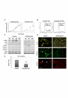
Inhibitory effect of LM-234ep conditioned medium on the proliferation of LM-234mf cells. LM-234mf and LM-234ep cells were seeded into 6-well plates at a density of 1 × 10⁴ cells/well and allowed to attach overnight. Thereafter, cells were incubated in the presence of LM-234ep CM (black circles), LM-234mf CM (black triangles), or culture medium (black squares) supplemented with 10% FBS. Cell numbers were counted at different time points, and expressed as mean ± SE. * P = 0.05, ** P = 0.0018 (Student's t Test). Assays were performed in triplicate, (A). LM-234mf cells were incubated with culture medium + 10% FBS (Control) or LM-234ep CM for 72 h. Cells were processed for DNA content, and cell cycle progression was analyzed by flow cytometry (B). LM-234mf cells were treated with culture medium (Control) or LM-234ep CM for the times indicated (top). Cells were collected, lysed, and analyzed by immunoblotting using antibodies specific for p-ERK, c-Jun, JunB, cyclins E, A, and D, and Cdk2. β-actin was used as a loading control (C). Immunofluorescence for c-Jun, Cyclin A and Cyclin D in LM-234mf cells. The nuclear localization of the proteins is shown in the control cells (white arrows). Magnification bar, 40 μm (D). AP-1 luciferase activity in LM-234mf cells co-transfected with AP-1-Luc and pRL-tk-luc and treated with LM-234ep CM or culture medium (Control). ** P = 0.005 (Student's t Test). The results shown are the mean ± S.D. of three experiments (E).
Figure 6
Figure 6
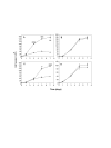
LM-234ep conditioned medium inhibits cell proliferation in cancer cells. B16-F10 (A and B), or HeLa (C and D) cells were seeded into 6-well plates at a density of 1 × 10⁴ cells/well and allowed to attach overnight. Cells were incubated with LM-234ep CM (black circles), LM-234mf CM (black triangles), or culture medium (black squares) containing 10% FBS (Control), and counted at different time points. Data are shown as mean ± SE. * P = 0.05, ** P = 0.0018, *** P = 0.0006, **** P < 0.0001 (Student's t Test). Assays were performed in triplicate.
Figure 7
Figure 7
Effects of LM-234ep conditioned medium on cell cycle and its regulatory proteins. B16-F10 and HeLa cells were treated with culture medium (Control) or LM-234ep CM for 48 h. Cell cycle progression was analyzed by flow cytometry (A). After the treatment for the indicated times (top) with LM-234ep CM or culture medium (Control), B16-F10 (B) and HeLa (D) cells were collected, lysed, and analyzed by immunoblotting using antibodies specific for cyclins E, A, and D, and Cdk2. β-actin was used as a loading control. Immunofluorescence for Cyclin A1 in B16-F10 (C), and HeLa (E) cells after 24 h incubation with culture medium or LM-234ep CM. Magnification bar, 40 μm.
Figure 8
Figure 8
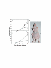
Inhibition of in vivo tumor growth by LM-234ep conditioned medium. B16-F10 (A) and HeLa (B) tumors growing in the right flank were injected with serum-free culture medium (black squares), whereas tumors in the left flank were injected with LM-234ep CM (black triangles). Tumors were measured and volumes were calculated as mean ± SE. * P = 0.018, ** P = 0.007, *** P = 0.004 (Student's t Test). Representative picture of a mouse showing inhibition of HeLa tumor growth by LM-234ep CM (arrow) as compared to culture medium treatment (contralateral flank), 4 weeks after tumor inoculation (C).

Similar articles

Cited by

References

    1. Jemal A, Siegel R, Ward E, Murray T, Xu J, Smigal C, Thun MJ. Cancer statistics, 2006. CA Cancer J Clin. 2006;56:106–130. - PubMed
    1. Ronnov-Jessen L, Petersen OW, Bissell MJ. Cellular changes involved in conversion of normal to malignant breast: importance of the stromal reaction. Physiol Rev. 1996;76:69–125. - PubMed
    1. Skobe M, Fusenig NE. Tumorigenic conversion of immortal human keratinocytes through stromal cell activation. Proc Natl Acad Sci U S A. 1998;95:1050–1055. doi: 10.1073/pnas.95.3.1050. - DOI - PMC - PubMed
    1. Bissell MJ, Radisky D. Putting tumours in context. Nat Rev Cancer. 2001;1:46–54. doi: 10.1038/35094059. - DOI - PMC - PubMed
    1. Ronnov-Jessen L, Petersen OW, Koteliansky VE, Bissell MJ. The origin of the myofibroblasts in breast cancer. Recapitulation of tumor environment in culture unravels diversity and implicates converted fibroblasts and recruited smooth muscle cells. J Clin Invest. 1995;95:859–873. - PMC - PubMed

Publication types

MeSH terms