

Agmatine protects retinal ganglion cells from hypoxia-induced apoptosis in transformed rat retinal ganglion cell line
- PMID: 17908330
- PMCID: PMC2089075
- DOI: 10.1186/1471-2202-8-81
Agmatine protects retinal ganglion cells from hypoxia-induced apoptosis in transformed rat retinal ganglion cell line
Retraction in
-
Retraction Note: Agmatine protects retinal ganglion cells from hypoxia-induced apoptosis in transformed rat retinal ganglion cell line.BMC Neurosci. 2023 Aug 14;24(1):42. doi: 10.1186/s12868-023-00814-3. BMC Neurosci. 2023. PMID: 37580650 Free PMC article. No abstract available.
Abstract
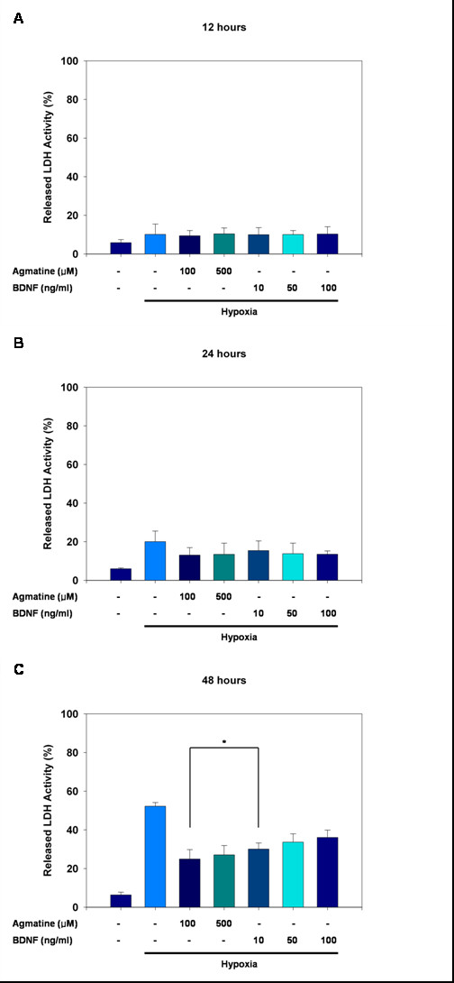
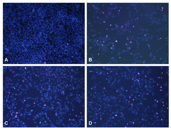
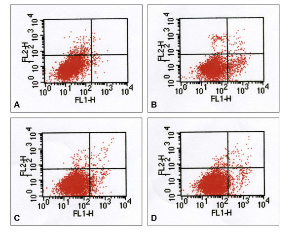
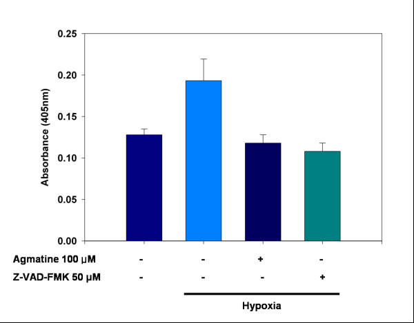
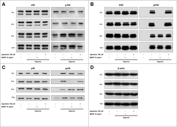
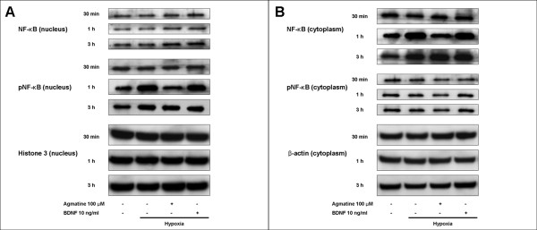
Background: Agmatine is an endogenous polyamine formed by the decarboxylation of L-arginine. We investigated the protective effects of agmatine against hypoxia-induced apoptosis of immortalized rat retinal ganglion cells (RGC-5). RGC-5 cells were cultured in a closed hypoxic chamber (5% O2) with or without agmatine. Cell viability was determined by lactate dehydrogenase (LDH) assay and apoptosis was examined by annexin V and caspase-3 assays. Expression and phosphorylation of mitogen-activated protein kinases (MAPKs; JNK, ERK p44/42, and p38) and nuclear factor-kappa B (NF-kappaB) were investigated by Western immunoblot analysis. The effects of agmatine were compared to those of brain-derived neurotrophic factor (BDNF), a well-known protective neurotrophin for retinal ganglion cells.
Results: After 48 hours of hypoxic culture, the LDH assay showed 52.3% cell loss, which was reduced to 25.6% and 30.1% when agmatine and BDNF were administered, respectively. This observed cell loss was due to apoptotic cell death, as established by annexin V and caspase-3 assays. Although total expression of MAPKs and NF-kappaB was not influenced by hypoxic injury, phosphorylation of these two proteins was increased. Agmatine reduced phosphorylation of JNK and NF-kappaB, while BDNF suppressed phosphorylation of ERK and p38.
Conclusion: Our results show that agmatine has neuroprotective effects against hypoxia-induced retinal ganglion cell damage in RGC-5 cells and that its effects may act through the JNK and NF-kappaB signaling pathways. Our data suggest that agmatine may lead to a novel therapeutic strategy to reduce retinal ganglion cell injury related to hypoxia.
Figures
Similar articles
-
Agmatine protects cultured retinal ganglion cells from tumor necrosis factor-alpha-induced apoptosis.Life Sci. 2009 Jan 2;84(1-2):28-32. doi: 10.1016/j.lfs.2008.10.006. Epub 2008 Oct 28. Life Sci. 2009. PMID: 18992261
-
Agmatine pretreatment protects retinal ganglion cells (RGC-5 cell line) from oxidative stress in vitro.Biocell. 2008 Dec;32(3):245-50. Biocell. 2008. PMID: 19181187
-
IGF-1 protects retinal ganglion cells from hypoxia-induced apoptosis by activating the Erk-1/2 and Akt pathways.Mol Vis. 2013 Sep 12;19:1901-12. eCollection 2013. Mol Vis. 2013. PMID: 24049436 Free PMC article.
-
Neurotrophin roles in retinal ganglion cell survival: lessons from rat glaucoma models.Exp Eye Res. 2009 Apr;88(4):808-15. doi: 10.1016/j.exer.2009.02.004. Epub 2009 Feb 14. Exp Eye Res. 2009. PMID: 19217904 Free PMC article. Review.
-
Neuroprotection by agmatine: Possible involvement of the gut microbiome?Ageing Res Rev. 2023 Nov;91:102056. doi: 10.1016/j.arr.2023.102056. Epub 2023 Sep 9. Ageing Res Rev. 2023. PMID: 37673131 Review.
Cited by
-
Beneficial effect of agmatine on brain apoptosis, astrogliosis, and edema after rat transient cerebral ischemia.BMC Pharmacol. 2010 Sep 6;10:11. doi: 10.1186/1471-2210-10-11. BMC Pharmacol. 2010. PMID: 20815926 Free PMC article.
-
Agmatine protects against zymosan-induced acute lung injury in mice by inhibiting NF-κB-mediated inflammatory response.Biomed Res Int. 2014;2014:583736. doi: 10.1155/2014/583736. Epub 2014 Aug 27. Biomed Res Int. 2014. PMID: 25243152 Free PMC article.
-
Exon 4-encoded sequence is a major determinant of cytotoxicity of apolipoprotein L1.Am J Physiol Cell Physiol. 2015 Jul 1;309(1):C22-37. doi: 10.1152/ajpcell.00384.2014. Epub 2015 Apr 29. Am J Physiol Cell Physiol. 2015. PMID: 25924622 Free PMC article.
-
The protective effect of melatonin on neural stem cell against LPS-induced inflammation.Biomed Res Int. 2015;2015:854359. doi: 10.1155/2015/854359. Epub 2015 Feb 1. Biomed Res Int. 2015. PMID: 25705693 Free PMC article.
-
TGF-β signaling is required for maintenance of retinal ganglion cell differentiation and survival.Neuroscience. 2011 Aug 25;189:123-31. doi: 10.1016/j.neuroscience.2011.05.020. Epub 2011 May 27. Neuroscience. 2011. PMID: 21664439 Free PMC article.
References
Publication types
MeSH terms
Substances
LinkOut - more resources
Full Text Sources
Other Literature Sources
Research Materials
Miscellaneous
