. 2007 Dec;27(23):8388-400.
doi: 10.1128/MCB.01493-07. Epub 2007 Oct 1.

Functional analysis of KSRP interaction with the AU-rich element of interleukin-8 and identification of inflammatory mRNA targets

Affiliations

Functional analysis of KSRP interaction with the AU-rich element of interleukin-8 and identification of inflammatory mRNA targets

Reinhard Winzen et al. Mol Cell Biol. 2007 Dec.

Abstract

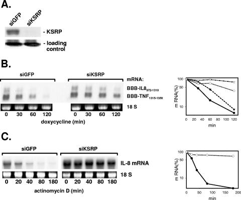
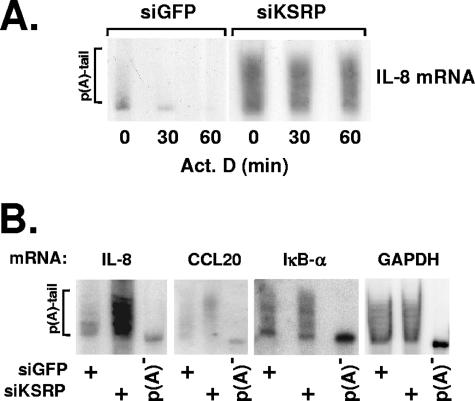
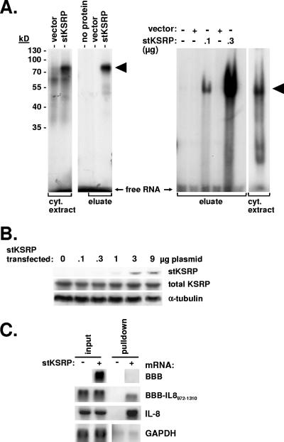
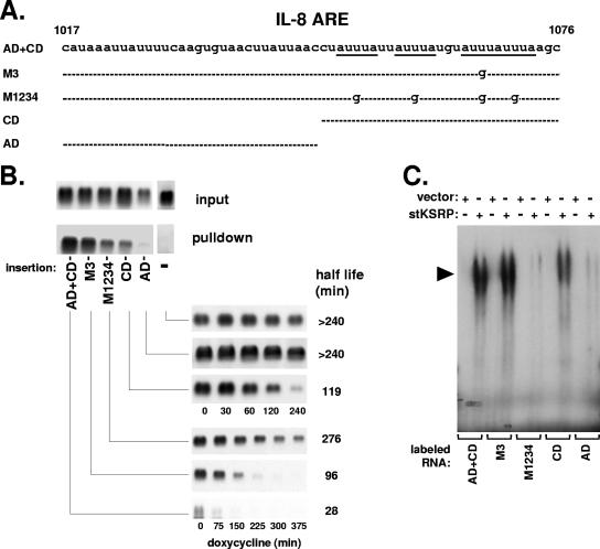
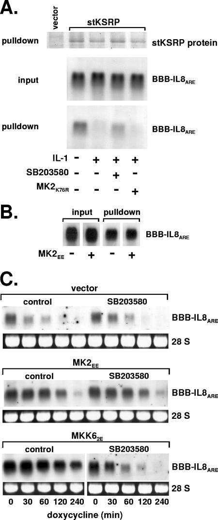
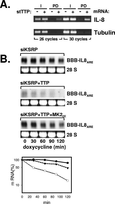
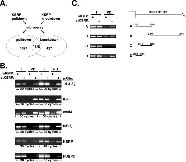
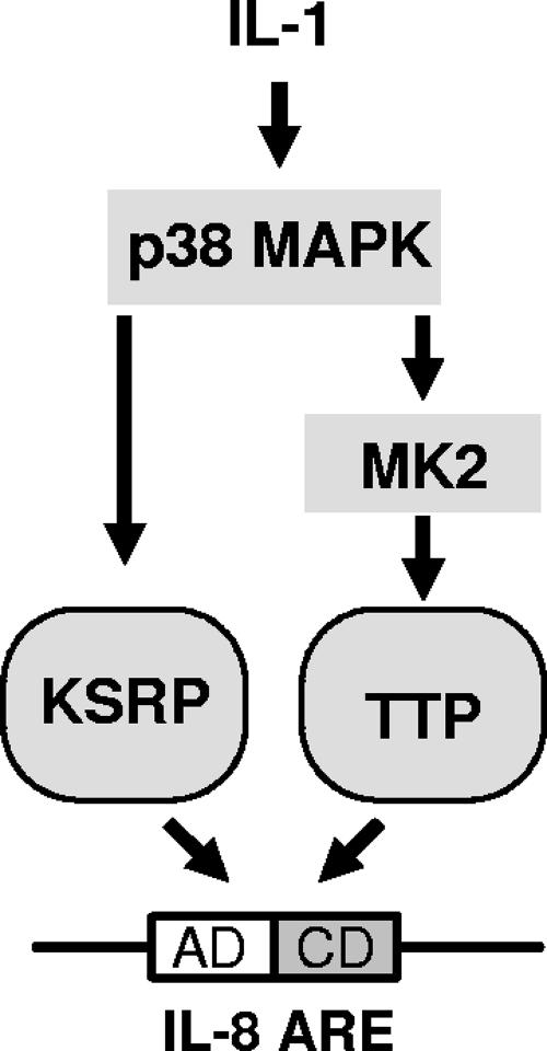
mRNA stability is a major determinant of inflammatory gene expression. Rapid degradation of interleukin-8 (IL-8) mRNA is imposed by a bipartite AU-rich element (ARE) in the 3' untranslated region (R. Winzen et al., Mol. Cell. Biol. 24:4835-4847, 2004). Small interfering RNA-mediated knockdown of the ARE-binding protein KSRP resulted in stabilization of IL-8 mRNA or of a beta-globin reporter mRNA containing the IL-8 ARE. Rapid deadenylation was impaired, indicating a crucial role for KSRP in this step of mRNA degradation. The two IL-8 ARE domains both contribute to interaction with KSRP, corresponding to the importance of both domains for rapid degradation. Exposure to the inflammatory cytokine IL-1 has been shown to stabilize IL-8 mRNA through p38 mitogen-activated protein (MAP) kinase and MK2. IL-1 treatment impaired the interaction of KSRP with the IL-8 ARE in a manner dependent on p38 MAP kinase but apparently independent of MK2. Instead, evidence that TTP, a target of MK2, can also destabilize the IL-8 ARE reporter mRNA is presented. In a comprehensive approach to identify mRNAs controlled by KSRP, two criteria were evaluated by microarray analysis of (i) association of mRNAs with KSRP in pulldown assays and (ii) increased amounts in KSRP knockdown cells. According to both criteria, a group of 100 mRNAs is controlled by KSRP, many of which are unstable and encode proteins involved in inflammation. These results indicate that KSRP functions as a limiting factor in inflammatory gene expression.


Figures

FIG. 1.
FIG. 1.
Evidence for a crucial role for KSRP in the control of IL-8 mRNA decay. HeLa cells were transfected with siRNA specific for KSRP or for GFP as a control (siKSRP or siGFP, respectively). (A) Western blot with antibodies against KSRP to control knockdown efficiency. Coomassie brilliant blue staining of an irrelevant protein is shown as a loading control. (B) Cells were cotransfected with plasmids for β-globin mRNAs containing destabilizing elements of IL-8 or of TNF-α mRNA. At the indicated times after stopping transcription with doxycycline (3 μg/ml), total RNA was isolated and analyzed by Northern blotting with a β-globin antisense probe. Ethidium bromide staining of the 18S rRNA is shown to allow comparison of the RNA amounts loaded. Results were quantified by a video analyzer system (amount of mRNA at the time of doxycycline addition [0 min] = 100%; circles, BBB-IL-8972-1310; triangles, BBB-TNF1315-1350; closed symbols, siGFP; open symbols, siKSRP). (C) Endogenous IL-8 mRNA was induced by incubating the cells for 2 h with IL-1α (2 ng/ml). mRNA half-life was determined after stopping transcription with actinomycin D (5 μg/ml). Northern blots were hybridized to an IL-8 antisense probe and results quantified as described for panel B.
FIG. 2.
FIG. 2.
Effect of KSRP knockdown on the poly(A) [p(A)] lengths of mRNAs. IL-1α was added to HeLa cells transfected with siRNA specific for KSRP or for GFP (siKSRP or siGFP, respectively). (A) After 2 h, transcription was inhibited by addition of actinomycin D (Act. D; 5 μg/ml). Total RNA was isolated at the indicated times thereafter, separated on a polyacrylamide gel, and analyzed by Northern blotting with an IL-8 antisense probe. (B) Polyacrylamide gel analysis of the indicated mRNAs from cells transfected with siRNAs and stimulated with IL-1α as described for panel A. Aliquots of the RNAs from cells transfected with siRNA against KSRP were subjected to in vitro deadenylation [p(A)].
FIG. 3.
FIG. 3.
Interaction of the IL-8 ARE with KSRP. (A) Radiolabeled in vitro-transcribed RNA corresponding to the IL-8 ARE was incubated alone (no protein), with cytoplasmic (cyt.) extracts from cells transfected with empty vector or with an stKSRP expression plasmid, or with eluate from Strep-tactin-agarose beads after incubation with the cytoplasmic extracts. Complexes were either separated by SDS-PAGE after UV cross-linking in vitro (left) or separated by nondenaturing gel electrophoresis (right). KSRP-RNA complexes are marked by arrowheads. (B) stKSRP expression plasmid was transfected in increasing amounts. Western blot detection of stKSRP was done with horseradish peroxidase-labeled Strep-tactin, and total KSRP and α-tubulin were detected with specific antibodies. (C) β-Globin reporter mRNAs without insertion (BBB) or with an IL-8 RNA fragment containing the ARE (BBB-IL-8972-1310) were coexpressed with stKSRP where indicated and the cells stimulated with IL-1α. Total RNA was prepared from cytoplasmic extract (input) and from eluate of Strep-tactin beads (pulldown). β-Globin and endogenous IL-8 and GAPDH mRNAs were detected by Northern blot analysis.
FIG. 4.
FIG. 4.
Participation of both domains of the IL-8 ARE in KSRP interaction and destabilization. (A) Scheme of the IL-8 ARE and derivatives assayed (CD, core domain; AD, auxiliary domain). (B) RNA isolated from cytoplasmic extract (input) and stKSRP pulldown was subjected to Northern blot analysis of β-globin reporter mRNAs without insertion (−), containing the complete IL-8 ARE (AD+CD) or mutants in which the third AUUUA motif (M3) or all four motifs (M1234) were changed into AUGUA, or containing a single domain as indicated. Also shown are the corresponding degradation kinetics of these mRNAs in HeLa cells (taken from reference with permission). (C) Complexes formed between purified stKSRP (0.4 μg) and radiolabeled RNA fragments corresponding to the complete IL-8 ARE or its derivatives were analyzed by nondenaturing gel electrophoresis. KSRP-RNA complexes are marked by arrowheads.
FIG. 5.
FIG. 5.
Effect of the p38 MAP kinase cascade on the interaction of KSRP with the IL-8 ARE. (A) β-globin reporter mRNA containing the IL-8 ARE was coexpressed with stKSRP and a negative interfering mutant of MK2 (MK2K76R) where indicated. The cells were incubated without or with IL-1α (2 ng/ml) for 30 min in the absence or presence of SB203580 (2 μM) added 15 min earlier. After pulldown of stKSRP, the protein was detected by SDS-PAGE and Coomassie brilliant blue staining, and associated β-globin-IL-8 ARE mRNA was detected by Northern blot analysis. (B) Pulldown of stKSRP and mRNA detection were performed as described for panel A for cells cotransfected with an expression vector for MK2EE as indicated. (C) Degradation kinetics of β-globin-IL-8 ARE mRNA in cells transfected with empty vector or plasmids for expression of constitutively active mutants of MK2 (MK2EE) or MKK6 (MKK62E) in the absence or presence of SB203580 (2 μM).
FIG. 6.
FIG. 6.
Interaction of TTP with the IL-8 ARE. (A) HeLa cells were transfected with a plasmid carrying stTTP or empty vector. Total RNA from cytoplasmic extract (I) or Strep-tactin pulldown (PD) was analyzed by RT-PCR, with primers specific for IL-8 mRNA or β-tubulin mRNA as a control. (B) HeLa cells were transfected with siRNA against KSRP, a plasmid carrying the β-globin-IL-8 ARE reporter RNA, and expression vectors for stTTP and MK2EE as indicated. The degradation kinetics of the reporter RNA was determined by Northern blot analysis and quantified as described for Fig. 1B. Closed circles, siKSRP; open circles, siKSRP plus TTP; triangles, siKSRP plus TTP plus MK2EE.
FIG. 7.
FIG. 7.
Interaction of KSRP with endogenous mRNAs. (A) HeLa cells were transfected with stKSRP or stGFP as a control for pulldown assays and with siRNA against KSRP or against GFP as a control for knockdown assays. After stimulation for 2 h with IL-1α, total RNA was isolated and subjected to microarray analysis as described in Materials and Methods. mRNAs associated with stKSRP in pulldown and increased in KSRP knockdown cells were identified. Details on mRNAs positive for both parameters (with ratios of >2 for signal intensities of stKSRP/stGFP and siRNA against KSRP/GFP) are presented in Table 1. (B) Enrichment of the indicated mRNAs in the stKSRP pulldown was confirmed by RT-PCR. (C) Input (I) and stKSRP pulldown (PD) samples from cells expressing β-globin mRNA with the indicated insertions of the KSRP 3′ UTR were analyzed for the respective mRNA by RT-PCR with β-globin mRNA-specific primers.
FIG. 8.
FIG. 8.
Scheme of IL-8 ARE-dependent control of mRNA stability. The two destabilizing proteins KSRP and TTP can interact with the IL-8 ARE and promote degradation. Activators of the p38 MAP kinase pathway, like IL-1, can induce stabilization by impairing the function of KSRP via p38 MAP kinase and of TTP via MK2 (for details, see Discussion).

Similar articles

Cited by

References

    1. Bakheet, T., B. R. Williams, and K. S. Khabar. 2006. ARED 3.0: the large and diverse AU-rich transcriptome. Nucleic Acids Res. 34:D111-D114. - PMC - PubMed
    1. Ben-Levy, R., S. Hooper, R. Wilson, H. F. Paterson, and C. J. Marshall. 1998. Nuclear export of the stress-activated protein kinase p38 mediated by its substrate MAPKAP kinase-2. Curr. Biol. 8:1049-1057. - PubMed
    1. Briata, P., S. V. Forcales, M. Ponassi, G. Corte, C. Y. Chen, M. Karin, P. L. Puri, and R. Gherzi. 2005. p38-dependent phosphorylation of the mRNA decay-promoting factor KSRP controls the stability of select myogenic transcripts. Mol. Cell 20:891-903. - PubMed
    1. Brook, M., C. R. Tchen, T. Santalucia, J. McIlrath, J. S. Arthur, J. Saklatvala, and A. R. Clark. 2006. Posttranslational regulation of tristetraprolin subcellular localization and protein stability by p38 mitogen-activated protein kinase and extracellular signal-regulated kinase pathways. Mol. Cell. Biol. 26:2408-2418. - PMC - PubMed
    1. Brooks, S. A., J. E. Connolly, and W. F. Rigby. 2004. The role of mRNA turnover in the regulation of tristetraprolin expression: evidence for an extracellular signal-regulated kinase-specific, AU-rich element-dependent, autoregulatory pathway. J. Immunol. 172:7263-7271. - PubMed

Publication types

MeSH terms

LinkOut - more resources