. 2007 Dec;75(12):5669-77.
doi: 10.1128/IAI.01096-07. Epub 2007 Oct 1.

Chlamydia trachomatis species-specific induction of ezrin tyrosine phosphorylation functions in pathogen entry

Affiliations

Chlamydia trachomatis species-specific induction of ezrin tyrosine phosphorylation functions in pathogen entry

Kena A Swanson et al. Infect Immun. 2007 Dec.

Abstract

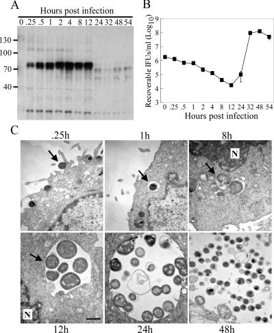
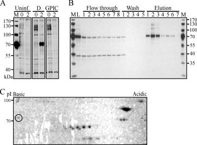
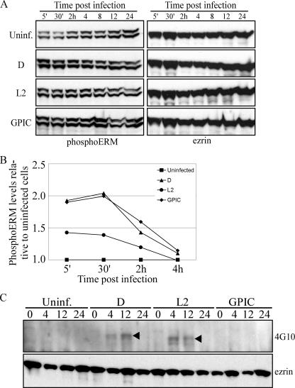
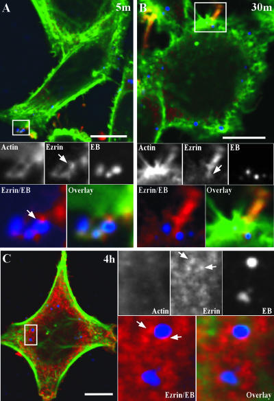
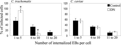
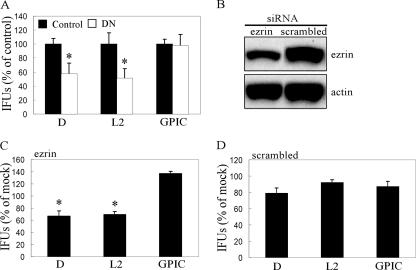
Chlamydia trachomatis is an obligate intracellular pathogen of humans that exhibits species-specific biological characteristics in its early interactions with host cells that are likely important to pathogenesis. One such characteristic is the tyrosine phosphorylation (Tyr-P) of an approximately 70-kDa polypeptide that occurs only after infection of mammalian cells by human strains. We sought to identify this protein because of its potential significance to the pathogenesis of human chlamydial infections. Using an immunoproteomic approach we identified the host protein ezrin, a member of the ezrin-radixin-moesin (ERM) protein family that serves as a physical link between host cell receptors and the actin cytoskeleton. Confocal microscopy studies showed colocalization of ezrin and actin at the tips and crypts of microvilli, the site of chlamydial attachment and entry, respectively. To demonstrate a functional role for ezrin we infected cells with a dominant-negative (DN) ezrin phenotype or treated cells with ezrin-specific small interfering RNA (siRNA). We found that both DN and siRNA-treated cells were significantly less susceptible to infection by human chlamydial strains. Moreover, we demonstrated that inhibition of infection in ezrin DN cells occurred at the stage of chlamydial entry. We hypothesize that the C. trachomatis-specific Tyr-P of ezrin might relate to an undefined species-specific mechanism of pathogen entry that involves chlamydial specific ligand(s) and host cell coreceptor usage.


Figures

FIG. 1.
FIG. 1.
The presence of Tyr-P correlates with the chlamydial developmental cycle. HeLa cells were infected with C. trachomatis D (MOI of ∼50). (A) Western blot analysis was performed on samples at the indicated times p.i. to examine phosphotyrosine levels using the 4G10 Ab. (B) Duplicate samples were harvested, and infectious chlamydiae were quantified by replating them on new HeLa cell monolayers. Infected cells were fixed 24 h p.i. and stained with an anti-LPS Ab, and inclusions were counted in triplicate wells. Error bars indicate the standard deviation. (C) HeLa cells were infected (MOI of ∼1) and fixed at the indicated times p.i. Coverslips were processed for TEM as described in Materials and Methods. Arrows in each image indicate the chlamydial EB at the early entry steps and subsequent trafficking of the early inclusion to the perinuclear region. Scale bar, 1 μm. N, nucleus.
FIG. 2.
FIG. 2.
Identification of ezrin as a host Tyr-P protein after C. trachomatis infection. (A) HeLa cells were infected with C. trachomatis D or GPIC (MOI of ∼1) or uninfected. Cells were harvested at 0 and 2 h p.i., and cleared lysates were separated by SDS-PAGE and immunoblotted with the 4G10 Ab. M, marker. (B) Tyr-P proteins were purified from C. trachomatis D-infected HeLa cells (MOI of ∼100) by 4G10 immunoaffinity chromatography. Purification efficiency is shown by 4G10 immunoblotting of equal volumes of collected fractions. M, marker; L, lysate. (C) Purified proteins were separated by 2D gel electrophoresis, followed by Coomassie blue staining. The circled protein spot indicates the ezrin protein identified by liquid chromatography-MS/MS.
FIG. 3.
FIG. 3.
C. trachomatis-specific ezrin Tyr-P is sustained to the mid-cycle of infection. (A) Ezrin Thr-P occurs independently of the infecting chlamydial strain. Cells remained uninfected or were infected with either C. trachomatis (D or L2) or GPIC (MOI of ∼0.5). Samples were harvested at the indicated times p.i. in Laemmli sample buffer, and Thr-P was determined by immunoblotting with an anti-phosphoERM Ab specific to the C-terminal Thr567. The same blots were stripped and reprobed for total ezrin. (B) Densitometry was performed on the blots shown in panel A. PhosphoERM levels were normalized to total ezrin levels within each sample. The graph shows the fold change in phosphoERM levels relative to uninfected cells, with values from uninfected cells set to 1. (C) Cells were infected as described above (MOI of ∼5), and ezrin was immunoprecipitated with an anti-rabbit ezrin Ab. The Tyr-P status was examined by Western blotting with the 4G10 Ab. The same blot was stripped and reprobed with the anti-ezrin Ab.
FIG. 4.
FIG. 4.
Ezrin recruitment to the EB entry site and early chlamydial inclusion. HeLa cells were infected with C. trachomatis D (MOI of ∼1). At 5 min (A) and 30 min (B) the cells were fixed and triple labeled by indirect immunofluorescence with anti-ezrin (red), anti-LPS (blue), and Alexa 488-conjugated phalloidin (green) for labeling of F-actin. (A) Confocal microscopy image showing ezrin and actin recruited to the tips of microvillus-like structures where three EBs are attached. The arrow indicates the ezrin-EB localization. Scale bar, 10 μm. (B) At 30 min p.i., ezrin is still seen associated with EBs during the entry process. The arrow indicates ezrin localization with EB in the crypt of the microvillus-like structure. Scale bar, 10 μm. (C) Ezrin localizes with early trafficking inclusions. HeLa cells were treated as in panels A and B except that the cells were fixed at 4 h p.i. Two trafficking early inclusions containing individual EBs are shown where ezrin is closely localized. Arrows indicate the punctate ezrin localization with the early inclusion. Scale bar, 2 μm.
FIG. 5.
FIG. 5.
Ezrin functions during C. trachomatis entry. Control or DN LLC-PK1 cells were infected with C. trachomatis D or GPIC (MOI of ∼0.01). Cells were fixed at 2 h p.i., and extracellular bacteria were stained with a MOMP-specific Ab. The cells were permeabilized, and the total bacteria were stained with an LPS-specific Ab. The number of internalized EBs was quantified and categorized according to number of intracellular EBs per cell. Only with C. trachomatis D infection was there a significant reduction in the number of EBs per infected cell. *, P < 0.002.
FIG. 6.
FIG. 6.
Ezrin-dependent C. trachomatis infection. (A) LLC-PK1 cells stably expressing the DN ezrin (DN, white bars) or control vector (Control, black bars) were infected with C. trachomatis (D or L2) or GPIC (MOI of ∼0.1). At 24 h p.i., the cells were fixed and stained with the anti-LPS Ab. Inclusions were quantified and normalized to infected control cells. In all experiments, control cells contained between approximately 200 to 300 inclusions across individual wells. Error bars indicate standard deviation. *, P < 0.005. (B) HeLa cells were transfected with ezrin-specific or scrambled ezrin siRNA. Ezrin protein levels were examined by Western blotting at 48 h posttransfection, the time of chlamydial infection. There was an ∼40% reduction in ezrin protein levels. (C and D) HeLa cells transfected with ezrin-specific (C) or scrambled (D) siRNA were infected 48 h posttransfection with C. trachomatis (D or L2) or GPIC (MOI of ∼0.01). Cells were fixed and stained 24 h p.i. as described above, and the inclusions were quantified. The values shown represent inclusion-forming units (IFUs) as a percentage of mock-transfected cells. *, P < 0.001.

Similar articles

Cited by

References

    1. Belland, R. J., G. Zhong, D. D. Crane, D. Hogan, D. Sturdevant, J. Sharma, W. L. Beatty, and H. D. Caldwell. 2003. Genomic transcriptional profiling of the developmental cycle of Chlamydia trachomatis. Proc. Natl. Acad. Sci. USA 100:8478-8483. - PMC - PubMed
    1. Birkelund, S., L. Bini, V. Pallini, M. Sanchez-Campillo, S. Liberatori, J. D. Clausen, S. Ostergaard, A. Holm, and G. Christiansen. 1997. Characterization of Chlamydia trachomatis L2-induced tyrosine-phosphorylated HeLa cell proteins by two-dimensional gel electrophoresis. Electrophoresis 18:563-567. - PubMed
    1. Birkelund, S., H. Johnsen, and G. Christiansen. 1994. Chlamydia trachomatis serovar L2 induces protein tyrosine phosphorylation during uptake by HeLa cells. Infect. Immun. 62:4900-4908. - PMC - PubMed
    1. Bretscher, A. 1983. Purification of an 80,000-dalton protein that is a component of the isolated microvillus cytoskeleton, and its localization in nonmuscle cells. J. Cell Biol. 97:425-432. - PMC - PubMed
    1. Caldwell, H. D., J. Kromhout, and J. Schachter. 1981. Purification and partial characterization of the major outer membrane protein of Chlamydia trachomatis. Infect. Immun. 31:1161-1176. - PMC - PubMed

Publication types

MeSH terms