. 2007 Dec;75(12):5788-97.
doi: 10.1128/IAI.00821-07. Epub 2007 Oct 1.

Phosphoinositide-3-kinase-dependent, MyD88-independent induction of CC-type chemokines characterizes the macrophage response to Toxoplasma gondii strains with high virulence

Affiliations

Phosphoinositide-3-kinase-dependent, MyD88-independent induction of CC-type chemokines characterizes the macrophage response to Toxoplasma gondii strains with high virulence

Chiang W Lee et al. Infect Immun. 2007 Dec.

Abstract

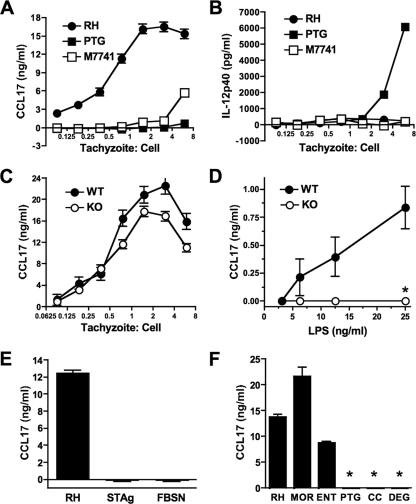
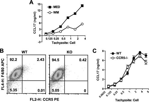
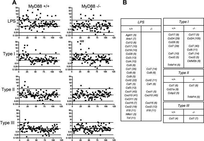
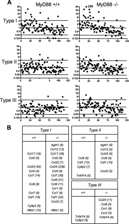
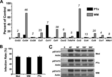
Chemokines play an important role in inflammation and infection due to their ability to recruit cells of innate and adaptive immunity. Here we examined mouse macrophage chemokine responses during intracellular infections with high- and low-virulence Toxoplasma gondii strains. The high-virulence type I strain RH induced a large panel of CC-type chemokines, whereas responses elicited by strains PTG (type II) and M7741 (type III) were much weaker. Strikingly, the T. gondii-induced chemokine response occurred independently of signaling through the Toll-like receptor adaptor MyD88. Instead, production of chemokines during infection was heavily dependent upon phosphoinositide-3-kinase signaling pathways. Because infection with type I strains such as RH results in an uncontrolled proinflammatory cytokine response, we hypothesize that this virulence phenotype is a consequence of early strong induction of chemokines by type I, but not type II or III, Toxoplasma strains.


Figures

FIG. 1.
FIG. 1.
Infection with Toxoplasma type I (RH), but not type II (PTG) or III (M7741), tachyzoites induces MyD88-independent CCL17 production. Bone marrow-derived macrophages were incubated for 18 h with RH, PTG, and M7741, and supernatants were collected for analysis of CCL17 (A) and IL-12p40 (B). (C) Wild-type (WT) and MyD88 knockout (KO) mice were infected with strain RH tachyzoites, and CCL17 levels were assessed 18 h later. (D) Wild-type and knockout cells stimulated with LPS. (E) Wild-type macrophages were incubated with live RH strain parasites (3:1 ratio of tachyzoites to cells), soluble tachyzoite lysate (STAg; 40 μg/ml), or cell-free supernatant from a culture of infected fibroblasts containing egressed parasites (FBSN; diluted 1:1). (F) Ability of RH, MOR, and ENT (type I strains) and PTG, CC, and DEG (type II strains) to induce CCL17 production. This experiment employed a 1.5:1 ratio of tachyzoites to cells. *, <60 pg/ml. The experiments were repeated two to four times with similar results.
FIG. 2.
FIG. 2.
Macrophage CCL17 response during Toxoplasma RH infection depends upon PI 3-kinase signaling. (A) Bone marrow-derived macrophages were preincubated for 60 min with wortmannin (50 ng/ml) and then infected with RH strain parasites. Supernatants were collected for ELISA 18 h later. (B) CCR5 expression in wild-type (WT) and CCR5 knockout (KO) macrophages. The mean fluorescence intensities were 8.1 and 7.3 for wild-type and knockout macrophages, respectively. (C) RH-induced CCL17 production in wild-type and CCR5 knockout macrophages. These experiments were repeated twice with the same results.
FIG. 3.
FIG. 3.
Chemokine response patterns during early T. gondii infection or LPS stimulation of wild-type and MyD88 knockout macrophages. Bone marrow-derived macrophages were stimulated with LPS (100 ng/ml) or infected with type I (RH), type II (PTG), or type III (M7741) tachyzoites, and samples were collected at 6 h postinfection for analysis. A 6:1 ratio of parasites to cells was used, yielding >80% infection. (A) Response patterns of 114 chemokine, chemokine receptor, and related genes. Values are expressed relative to those for nonstimulated cells. The horizontal line defines 3 standard deviations from the mean for all genes for the sample showing the least amount of change (type III infection of MyD88−/− macrophages). (B) Identities of genes upregulated by parasites and LPS. Numbers in parentheses indicate x-fold increases relative to levels in nonstimulated cells.
FIG. 4.
FIG. 4.
Chemokine response patterns during late T. gondii infection of wild-type and MyD88−/− macrophages. The experiment was set up as described in the legend to Fig. 1, with samples collected at 18 h postinfection.
FIG. 5.
FIG. 5.
Inhibitory effects of blocking Gi protein and PI 3-kinase signaling on type I Toxoplasma infection. (A) Macrophages were preincubated for 2 h with pertussis toxin (PTx) or wortmannin (WM) and then infected with RH strain parasites at a 6:1 ratio of tachyzoites to cells. Samples were collected at 6 h postinfection for analysis. Values are expressed relative to responses in the absence of inhibitors. The x-fold increase of each gene in the absence of inhibitors is shown above the bars for each gene. (B) Infection rates of macrophages in the presence and absence (Med) of inhibitors. Cells were preincubated with 50 ng/ml each of wortmannin and pertussis toxin, and type I infection was carried out at a 6:1 ratio of parasites to cells. (C) Phosphorylation of STAT3 in the presence and absence of pertussis toxin and wortmannin. Cells were pretreated with inhibitors and infected with RH tachyzoites, and samples were collected for Western blot analysis at the indicated time points.
FIG. 6.
FIG. 6.
Chemokine response patterns during in vivo infections with Toxoplasma strain types I, II, and III. Tachyzoites (106) were inoculated i.p. into mice (three per group), and peritoneal cells were collected at 6 and 18 h postinjection. RNAs were prepared and chemokine gene expression analyzed relative to that in cells from mice injected with PBS alone. (A) Patterns of chemokine, chemokine receptor, and related genes expressed during infection. (B) Identities of genes upregulated during infection. Numbers in parentheses indicated x-fold upregulation relative to the levels in noninfected control animals.

Similar articles

Cited by

References

    1. Aliberti, J., C. Reis e Sousa, M. Schito, S. Hieny, T. Wells, G. B. Huffnage, and A. Sher. 2000. CCR5 provides a signal for microbial induced production of IL-12 by CD8α+ dendritic cells. Nat. Immunol. 1:83-87. - PubMed
    1. Aliberti, J., J. G. Valenzuela, V. B. Carruthers, S. Hieny, J. Andersen, H. Charest, C. Reis e Sousa, A. Fairlamb, J. M. Ribeiro, and A. Sher. 2003. Molecular mimicry of a CCR5 binding-domain in the microbial activation of dendritic cells. Nat. Immunol. 4:485-490. - PubMed
    1. Allen, S. J., S. E. Crown, and T. M. Handel. 2007. Chemokine: receptor structure, interactions, and antagonism. Annu. Rev. Immunol. 25:787-820. - PubMed
    1. Bjorkbacka, H., K. A. Fitzgerald, F. Huet, X. Li, J. A. Gregory, M. A. Lee, C. M. Ordija, N. E. Dowley, D. T. Golenbock, and M. W. Freeman. 2004. The induction of macrophage gene expression by LPS predominantly utilizes MyD88-independent signaling cascades. Physiol. Genomics 19:319-330. - PubMed
    1. Bonecchi, R., G. Bianchi, P. P. Bordignon, D. D'Ambrosio, R. Lang, A. Borsatti, S. Sozzani, P. Allavena, P. A. Gray, A. Mantovani, and F. Sinigaglia. 1998. Differential expression of chemokine receptors and chemotactic responsiveness of type 1 T helper cells (Th1s) and Th2s. J. Exp. Med. 187:129-134. - PMC - PubMed

Publication types

MeSH terms

LinkOut - more resources